Indexed In
- Open J Gate
- Academic Keys
- RefSeek
- Hamdard University
- EBSCO A-Z
- OCLC- WorldCat
- Publons
- Euro Pub
- Google Scholar
- SHERPA ROMEO
Useful Links
Share This Page
Journal Flyer

Open Access Journals
- Agri and Aquaculture
- Biochemistry
- Bioinformatics & Systems Biology
- Business & Management
- Chemistry
- Clinical Sciences
- Engineering
- Food & Nutrition
- General Science
- Genetics & Molecular Biology
- Immunology & Microbiology
- Medical Sciences
- Neuroscience & Psychology
- Nursing & Health Care
- Pharmaceutical Sciences
Case Report - (2023) Volume 0, Issue 0
'United We Stand": A Rare Case of Non-Incorporation of the Common Pulmonary Vein into Left Atrium
Geetha Sulochana Gopidas* and Mrudula ChandrupatlaReceived: 21-Mar-2023, Manuscript No. JVMS-23-20226; Editor assigned: 24-Mar-2023, Pre QC No. JVMS-23-20226 (PQ); Reviewed: 07-Apr-2023, QC No. JVMS-23-20226; Revised: 14-Apr-2023, Manuscript No. JVMS-23-20226 (R); Published: 24-Apr-2023, DOI: 10.35248/2329-6925.23.S15.512
Abstract
Purpose: Anatomic variations of left atrium that are commonly observed and reported are the occurrence of a common right or left pulmonary venous ostia though a solitary ostium for a common pulmonary vein on the left atrial wall in an adult cadaveric heart has hardly been reported before.
Method: A heart with a single ostium for PV was observed in the specimen collection of the gross anatomy lab. The organ was studied for identifying all external and internal features.
Results: A very rare case of a Solitary Pulmonary ostium on the posterior aspect of Left Atrium in an otherwise normal heart was observed.
Conclusion: This abnormal situation occurs when the solitary pulmonary vein which develops from an out-pouching of the primitive atrial chamber develops and its primary divisions fail to get incorporated into the left atrial wall as expected during development. Variant pulmonary veins have been reported as ectopic trigger spots for atrial fibrillation. This knowledge would benefit the radiologists and cardiovascular surgeons in this regard.
Keywords
Solitary pulmonary vein; Common pulmonary vein; Single pulmonary vein; Variant pulmonary veins; Pulmonary ostia
Keywords
Solitary pulmonary vein; Common pulmonary vein; Single pulmonary vein; Variant pulmonary veins; Pulmonary ostia
Introduction
Two pairs of right and left Pulmonary Vein (PV), superior and inferior drain into the Left Atrium (LA) from the corresponding side of the lungs in 82% of the population [1]. This is considered as normal. When the number of pulmonary veins draining into the LA varies from the normal, it is classified as abnormal numbers of PV [2,3]. Pulmonary veins are classified as anomalous only if one or all pulmonary veins abnormally connect to systemic veins instead of the left atrium. Until late 20th century, variations in the number and draining pattern of pulmonary veins reported were rare [4]. Recently with the advent of imaging technology and rise in research few more authors have reported variations in the number of pulmonary veins and their ostia in left atrium [2,5,6]. Yet, a Solitary/Single PV opening into the posterior wall of left atrium has not been reported in adults. In a fetal study where three cases of atresia of common PV has been reported in newborns were emergency situations and fatal [7]. Here we report a case of single ostia on the left atrium in an adult for drainage of a single PV.
Case Presentation
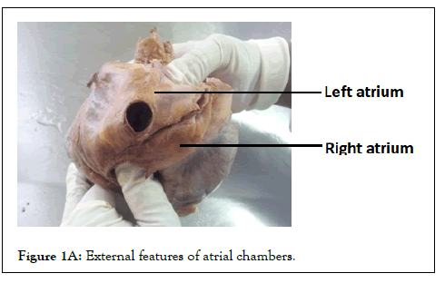
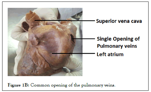
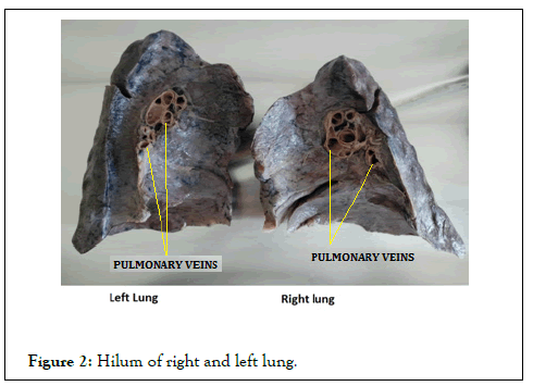
Among the specimen collection in the gross anatomy lab, we observed a heart with a single ostium for PV in the posterior left atrial wall, which had been dissected from a 70 yr old male cadaver (Figure 1A). Both atrial chambers, septal walls, and valves were normal. The right and left pulmonary veins were joining to form a common vein that drained to the posterior aspect of left atrium (Figure 1B). The hilum of the lungs did not show any variations and the pulmonary veins are normal (Figure 2).

Figure 1A: External features of atrial chambers.

Figure 1B: Common opening of the pulmonary veins.

Figure 2: Hilum of right and left lung.
Embryology
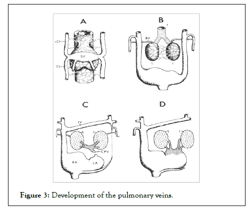
Development of pulmonary veins: Primitive pulmonary veins originate from venous plexus of splanchnic mesoderm around the developing lung buds at approximately 26 days. They communicate with the systemic cardinal and umbilico-vitelline veins. At 27 to 29 days, an out-pouching/an evagination develops from the posterosuperior wall of the primitive left atrium. As this pouch elongates it connects pulmonary venous networks to the heart tube near the sinu-atrial junction using the regressing dorsal mesocardium as portal of cardiac entry and is called the Solitary Pulmonary Vein (SPV) aka Common Pulmonary Vein (CPV). With the development of muscular septum primum, this along with the ventral proliferation of extracardiac mesenchyme from the dorsal mesocardium positions the CPV eventually into the left atrium occurs as shown in Figure 3. As the systemic veins regress they get separated from the walls of the CPV. The CPV and its first and second divisions become incorporated into left atrium by a morphologically asymmetric process. Thus four pulmonary veins Right Superior, Right Inferior, Left Superior and Left Inferior are eventually draining independently into the left atrium [8,9]. Defects in the formation of the posterior left atrial wall by absorption of the embryonal SPV and its divisions is the determinant factor that causes the anatomical variations in this area [9]. Partial incorporation of the CPV or the pulmonary veins into the left atrium can result in an abnormal number of pulmonary veins on right or left side. Total anomalous pulmonary venous return results when atresia of the Solitary Pulmonary Vein occurs before the separation of the pulmonary and splanchnic venous plexuses, detaching the pulmonary venous system from left atrium. Stenosis of the common pulmonary vein can result in cor-triatriatum [2,3].

Figure 3: Development of the pulmonary veins.
Results and Discussion
A Common Rt. PV or a Common Lt. PV, or a third Right PV are the frequent variants reported in the Pulmonary Venous System. A Single PV draining into the left atrium has been rarely reported [2,10,11]. A study by Narasipuram, et al. and Jongbloed, et al. compares the variations reported in a few previous studies and found that variations occur more commonly in the Right Pulmonary Vein than Left. These anomalies were most often incidental findings observed on imaging studies or cadaveric studies [11,12]. Persistence of the SPV presenting as a separate atretic chamber and lacking any connection with atrium has been reported as a very rare condition that requires immediate recognition and surgical intervention [13]. Knowing about the incidence and tracing the origin of these anomalous veins has great clinical relevance as it could be misread as an arteriovenous malformation or could be life threatening if noticed only intraoperatively [14].
Clinical significance
In Abnormal pulmonary veins are major sources of atrial (ectopic) trigger points for initiating frequent paroxysms of atrial fibrillation. Anomalies of PV are often seen in patients with Atrial Fibrillation [15]. Cryoballoon (CB)-guided Pulmonary Vein Isolation (PVI) is an established treatment for Atrial Fibrillation (AF) Variant PV anatomy seems to be predictive for AF recurrence [16].
Conclusion
Non-incorporation of the embryological Solitary Pulmonary Vein with its tributaries into the posterior left atrial wall and their persistence in an adult cadaveric specimen as reported in our case is a very rare occurrence. Variations in number of pulmonary veins are often detected in Atrial Fibrillation patients and may be associated with enlarged diameters of Left Atrial and Pulmonary Venous ostia. This knowledge would benefit the radiologists and cardiovascular surgeons in early diagnosis and prevention of Atrial Fibrillation.
Statements and Declarations
The work reported has not been published before; and it is not under consideration for publication anywhere else; its publication has been approved by all co-authors, as well as by the responsible authorities-tacitly or explicitly-at the institute where the work has been carried out. The publisher will not be held legally responsible should there be any claims for compensation.
Competing Interests and Funding
There are no competing interests that are directly or indirectly related to this work submitted for publication. No funds have been received for the same.
Compliance with Ethical Standards
This research involves dissected human cadaveric organ and is exempted from requiring ethics approval by the Institutional Ehics Committee.
References
- Cronin P, Kelly AM, Desjardins B, Patel S, Gross BH, Kazerooni EA, et al. Normative analysis of pulmonary vein drainage patterns on multi-detector CT with measurements of pulmonary vein ostial diameter and distance to first bifurcation. Acad Radiol. 2007;14(2):178-188.
[Crossref] [Google Scholar] [Pub Med]
- Healey Jr JE. An anatomic survey of anomalous pulmonary veins: Their clinical significance. J Thorac Surg. 1952;23(5):433-444.
[Crossref] [Google Scholar] [Pub Med]
- Neill CA. Development of the pulmonary veins: With reference to the embryology of anomalies of pulmonary venous return. Pediatrics. 1956;18(6):880-887.
[Crossref] [Google Scholar] [Pub Med]
- Alfke H, Wagner HJ, Klose KJ. A case of an anomalous pulmonary vein of the right middle lobe. Cardiovasc Intervent Radiol. 1995;18:406-409.
[Crossref] [Google Scholar] [Pub Med]
- Marom EM, Herndon JE, Kim YH, McAdams HP. Variations in pulmonary venous drainage to the left atrium: Implications for radiofrequency ablation. Radiology. 2004;230(3):824-829.
[Crossref] [Google Scholar] [Pub Med]
- Kaur A, Sharma A, Sharma M. Variations in the number of pulmonary veins draining into left atrium and its clinical significance. Inter J Med Dent Sci. 2017;6:1413-1416.
[Crossref] [Google Scholar]
- Perez M, Kumar TS, Briceno-Medina M, Alsheikh-Ali M, Sathanandam S, Knott-Craig CJ. Common pulmonary vein atresia: Report of three cases and review of the literature. Cardiol Young. 2016;26(4):629-635.
[Crossref] [Google Scholar] [Pub Med]
- Nathan H, Eliakim M. The junction between the left atrium and the pulmonary veins: An anatomic study of human hearts. Circulation. 1966;34(3):412-422.
[Crossref] [Google Scholar] [Pub Med]
- Reller MD, McDonald RW, Gerlis LM, Thornburg KL. Cardiac embryology: Basic review and clinical correlations. J Am Soc Echocardiogr. 1991;4(5):519-532.
[Crossref] [Google Scholar] [Pub Med]
- Hidvegi RS, Lapin J. Anomalous bilateral single pulmonary vein demonstrated by a 3-dimensional reconstruction of helical computed tomographic angiography: Case report. Can Assoc Radiol J. 1998;49(4):262-265.
- Narasipuram A, Chandraputula M, Fatima H. Anatomic study of number of pulmonary veins draining into the left atrium and their variations. J Med Sci Health. 2020;6(2):43-45.
- Jongbloed MR, Dirksen MS, Bax JJ, Boersma E, Geleijns K, Lamb HJ, et al. Atrial fibrillation: Multi-detector row CT of pulmonary vein anatomy prior to radiofrequency catheter ablation-initial experience. Radiology. 2005;234(3):702-709.
[Crossref] [Google Scholar] [Pub Med]
- Khonsari S, Saunders PW, Lees MH, Starr A. Common pulmonary vein atresia: Importance of immediate recognition and surgical intervention. J Thorac Cardiovasc Surg. 1982;83(3):443-448.
[Crossref] [Google Scholar] [Pub Med]
- Rasheed HA, Reinking BE. Anomalous single pulmonary venous trunk. Avicenna J Med. 2012;2(1):12-14.
[Crossref] [Google Scholar] [Pub Med]
- Skowerski M, Wozniak-Skowerska I, Hoffmann A, Nowak S, Skowerski T, Sosnowski M, et al. Pulmonary vein anatomy variants as a biomarker of atrial fibrillation-CT angiography evaluation. BMC Cardiovasc Disord. 2018;18:1-7.
[Crossref] [Google Scholar] [Pub Med]
- Guckel D, Lucas P, Isgandarova K, Hamriti ME, Bergau L, Fink T, et al. Impact of pulmonary vein variant anatomy and cross-sectional orifice area on freedom from atrial fibrillation recurrence after cryothermal single-shot guided pulmonary vein isolation. J Interv Card Electrophysiol. 2022;65(1):251-260.
[Crossref] [Google Scholar] [Pub Med]
Citation: Gopidas GS, Chandrupatla M (2023) ‘United We Stand”: A Rare Case of Non-Incorporation of the Common Pulmonary Vein into Left Atrium. J Vasc Surg. S15:512.
Copyright: © 2023 Gopidas GS, et al. This is an open-access article distributed under the terms of the Creative Commons Attribution License, which permits unrestricted use, distribution, and reproduction in any medium, provided the original author and source are credited.

