Indexed In
- Open J Gate
- Academic Keys
- RefSeek
- Hamdard University
- EBSCO A-Z
- OCLC- WorldCat
- Publons
- Euro Pub
- Google Scholar
- SHERPA ROMEO
Useful Links
Share This Page
Journal Flyer

Open Access Journals
- Agri and Aquaculture
- Biochemistry
- Bioinformatics & Systems Biology
- Business & Management
- Chemistry
- Clinical Sciences
- Engineering
- Food & Nutrition
- General Science
- Genetics & Molecular Biology
- Immunology & Microbiology
- Medical Sciences
- Neuroscience & Psychology
- Nursing & Health Care
- Pharmaceutical Sciences
Case Report - (2020) Volume 8, Issue 2
True Brachial Artery Aneurysm in Non-smoker Woman
Vu Hai-Minh1, Hoang Nang-Trong2 and Sy Duong-Quy3*2Department of Ophthalmology, Thai Binh University of Medicine and Pharmacy, Thai Binh, Vietnam
3Penn State Medical College, Hershey Medical Center, Pennsylvania, USA
Received: 18-Jan-2020 Published: 20-Feb-2020, DOI: 10.35248/2329-6925.20.8.386
Abstract
Aneurism is a stretch, convexity and sacs in artery which is caused by abnormally congenital connective tissue, infection, arterial inflammation or artery wall trauma. True brachial artery aneurysm is relatively rare. There are only some cases that have been reported previously in medical literature. Brachial artery aneurysm might be asymptomatic or revealed by a brachial mass with beating pulse or peripheral ischemia. The diagnosis usually relies on ultrasound and arterial scan or magnetic resonance imaging (MRI). The main treatment is the surgery which allows cutting the aneurysm and grafted with a part of great saphenous vein by two end-to-end anastomoses. We present a case report of true aneurysm of brachial artery in a 40 years old woman who was treated in Thai Binh Medical University Hospital.
Keywords
Brachial artery aneuryms; Aneuryms; Saphenous vein; Vascular surgery
Introduction
Brachial aneurysm is actually relatively rare. Until now, there are only a few cases that have been reported in the medical literature previously. The main clinical symptom is usually a pulsatile swelling on the arm that might be accidentally discovered or hospitalized with the symptoms of thrombotic anemia. We present here a real case of true brachial aneurysm of a 40 years old female farmer who was treated with a surgery at Thai Binh Medical University Hospital - Vietnam in December 2014. During 5-years followup in the outpatient department, there was not any symptom of aneurysm recurrence or other complications.
Case Report
A 40-years old and non-smoker woman who was admitted in our clinic with a swollen mass in the medial side of her arm which she had been carried for 9 years. Her medical history was negative for diabetes mellitus, hypertension or cardiovascular diseases. She had not any history of trauma or medical procedure in her arm. In her family, there was not any person who had the same problem with aneurysm. The clinical examination revealed a soft swelling of 3x2 cm diameter located in the third of the front-inner of right arm and on the tract of the brachial artery. This soft mass was pulsed synchronizing with right brachial artery beating. There was no evidence of anemia, pain, numbness, paresthesia and murmur at auscultation. Dropper’s examination and CT angiography showed an aneurysm with 17 mm long and 12 mm of the largest for horizontal diameter and 6 mm of the aneurysm’s neck diameter (Figure 1).
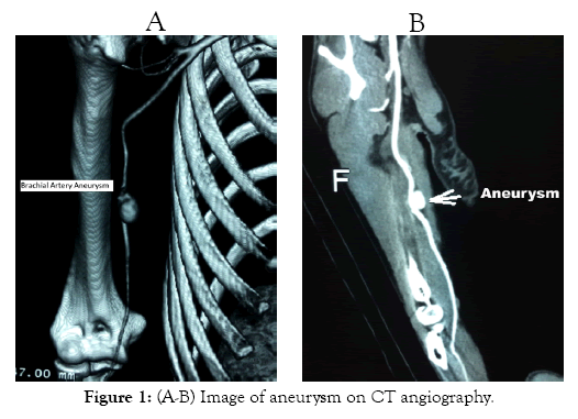
Figure 1: (A-B) Image of aneurysm on CT angiography.
This patient underwent brachial plexus block anesthesia and surgical treatment. A resection and anastomosis was performed with 8 cm at the medial third of the right arm. During her aneurysm operation, we found a mass of 2,2 x 1,7 cm size and it was then resected (Figure 2). The lesion was sutured with the graft with of saphenous to the right brachial artery (Figure 3). After operation, Heparin 7500 UI was injected intravenously by electric syringe for 48 hours to the patient and then a dose of Aspirin 100mg per day was used for 30 days. Clinical examination after five days of operation showed arteries at right elbow and radial artery pulsed worked well; the circuit turned right elbow had a good beating; arterial Doppler demonstrated the graft connection was circulated well (Figure 4). The patient was discharged six days after surgery. Re-examination and CT angiography 30 day postoperation showed that anastomotic stenosis did not happen (Figure 5).
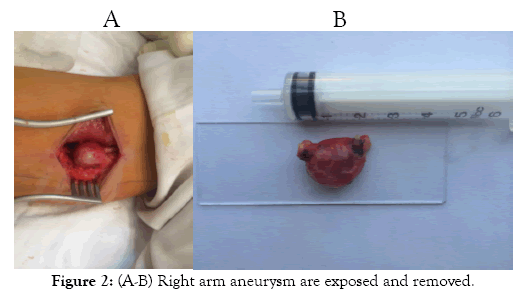
Figure 2: (A-B) Right arm aneurysm are exposed and removed.
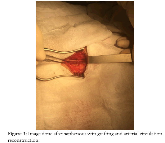
Figure 3: (A-B) Right arm aneurysm are exposed and removed.
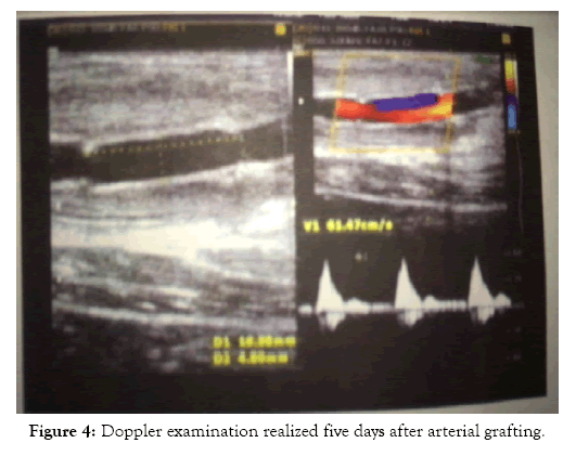
Figure 4: Doppler examination realized five days after arterial grafting.
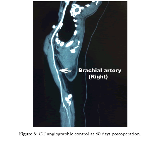
Figure 5: CT angiographic control at 30 days postoperation.
Discussion
Brachial artery aneurysm is rare in surgical practice. There have been very few reports on these aneurysms which threaten the upper limbs because of embolism in brachial vessels. Prompt diagnosis and treatment become necessary to minimize the complication rate and serious long-term effect. The mechanism of aneurysm formation is alleged to be the compression of arterial wall, producing contusion of the media and subsequent weakness of the wall that gradually transforms into fusiform dilatation [1]. There are two types of aneurysms: true aneurysms and pseudo-aneurysms of the brachial artery. The pseudo-aneurysm might be give a complication of cardiac catheterization performed for congenital heart disease, arterial and venous puncture for the evaluation of blood gases, and invasive arterial monitoring [2]. True aneurysm might be caused by arteriosclerotic, congenital, and metabolic disorders [3]. It might be associated with other diseases such as Kawasaki's syndrome, Buerger's disease, Kaposi's sarcoma, and cystic adventitial disease [4]. In our case, the patient did not have any of the previously aforementioned diseases and other diseases of thromboembolic origin.
The initial diagnosis may be made by clinic examination [5]. Most cases present as an asymptomatic pulsatile swelling. It might be presented with complications such as distal thromboembolism, forearm or hand ischemia, rupture, hemarthrosis, adjacent nerve irritation, paresthesia, limited elbow mobility, skin ulceration, and secondary infection [6]. Misdiagnosis of a ganglion remains the pitfall [7]. Other differential diagnosis includes pulsating tumors (such as bone sarcoma and osteoclastoma), anteriovenous malformation lymphadenopathy, lipoma, hematoma and abscess, etc… [8]. The diagnosis should be confirmed by a duplex ultrasonography, displays the arterial blood flow into the aneurysm. Computed tomography or magnetic resonance angiography might also be useful test supporting the diagnosis [6]. In spite of all abovementioned tests, the gold standard test is a selective upper extremity arteriography. However, the first choices in investigation are often color Doppler ultrasonography and subtraction image angiography [2]. The defect in the artery is usually small and easily identified by a transient release of the proximal clamp [5]. In the present case report, our patient was examined by Doppler ultrasonography and CT angiography. The close observation, thrombin injection, ultrasound-guided compression, and operative repair are the management options.
Because the brachial artery aneurysm is a rare pathology worldwide, the exact surgical procedure and definite treatment option for true upper extremity artery aneurysm has not been established [1,4,6]. Ultrasound-guided compression combined with injection of thrombin, radiologic intervention and surgical resection could be considered as one of the therapeutic modalities. Ultrasoundguided compression and thrombin injection have the advantages of a non-invasive procedure, but significant disadvantages such as pain and less efficacy in anticoagulation patients are usually associated with it [5]. Radiologic intervention for the brachial artery aneurysm might not be possible or not advisable around the elbow joint. A covered stent may be technically feasible but certainly not indicated in an area developing such flexion amplitude [1-6]. There are not many reports of brachial artery aneurysm that compares the nonsurgical and surgical treatment; therefore, its treatment is controversial. It should be considered that the best therapeutic option for brachial artery aneurysm might be the operative repair and it should be performed without any delay in order to prevent upper extremity ischemic sequelae because many authors have reported the surgical resection as the therapeutic modality [7,8]. When surgical resection is performed, it is important that the aneurysm wall with the changes should be resected with adequate resection margins. Then, the autologous reversed saphenous vein graft interpositioning could be performed. In our case report, the aneurysmectomy has been performed and reversed saphenous vein graft has been used for the arterial reconstruction because our patients had no a vascular structure permitting conducive to endto- end anastomosis.
Conclusion
Brachial artery aneurysm is rare in medical literature. Aneurysm might be the cause of thrombosis which threatens patient life with extremity ischemia. The amputation may be avoided for patients with early diagnosis and surgical removal technique. Our patient was discharged with successful results due to the timely detection and tight cooperation of multidisciplinary specialties.
REFERENCES
- Ho PK, Weiland AJ, McClinton MA, Wilgis EF. Aneurysms of the upper extremity. J Hand Surg Am. 1987;12:39-46.
- Schunn CD, Sullivan TM. Brachial arteriomegaly and true aneurysmal degeneration: case report and literature review. Vasc Med. 2002;7:25-27.
- Malt S. An arteriosclerotic aneurysm of the hand. Arch Surg. 1978;113:762-763.
- Hudorović N, Lovričević I, Franjić DB, Brkić P, Tomas D. True aneurysm of brachial artery. Wien Klin Wochenschr. 2010;122:588-591.
- Chemla E, Nortley M, Morsy M. Brachial artery aneurysms associated with arteriovenous access for hemodialysis. Semin Dial. 2010;23:440-444.
- Tetik O, Ozcem B, Calli AO, Gurbuz A. True brachial artery aneurysm. Tex Heart Inst J. 2010;37:618-619.
- Schunn CD, Sullivan TM. Brachial arteriomegaly and true aneurysmal degeneration: case report and literature review. Vasc Med. 2002;7(1):25-27.
- Ghazi MA, Khan AM, Akram Y, Cheema MA. Brachial artery aneurysm. Japan Med Assoc J. 2006;49(4):173.
Citation: Hai-Minh V, Nang-Trong H, Duong-Quy S (2020) True Brachial Artery Aneurysm in Non-smoker Woman. J Vasc Med Surg 8:386. doi: 10.35248/2329-6925.20.8.386
Copyright: © 2020 Hai-Minh V, et al. This is an open-access article distributed under the terms of the Creative Commons Attribution License, which permits unrestricted use, distribution, and reproduction in any medium, provided the original author and source are credited.

