Citations : 2345
Dentistry received 2345 citations as per Google Scholar report
Indexed In
- Genamics JournalSeek
- JournalTOCs
- CiteFactor
- Ulrich's Periodicals Directory
- RefSeek
- Hamdard University
- EBSCO A-Z
- Directory of Abstract Indexing for Journals
- OCLC- WorldCat
- Publons
- Geneva Foundation for Medical Education and Research
- Euro Pub
- Google Scholar
Useful Links
Share This Page
Journal Flyer

Open Access Journals
- Agri and Aquaculture
- Biochemistry
- Bioinformatics & Systems Biology
- Business & Management
- Chemistry
- Clinical Sciences
- Engineering
- Food & Nutrition
- General Science
- Genetics & Molecular Biology
- Immunology & Microbiology
- Medical Sciences
- Neuroscience & Psychology
- Nursing & Health Care
- Pharmaceutical Sciences
Research - (2019) Volume 9, Issue 5
The Evaluation of the Prevalence and Intensity of Dental Caries in β-thalassemia Major Patients
Shadlinskaya R.V.* and Zeynalova G.K.Received: 11-Jun-2019 Published: 25-Jul-2019, DOI: 10.35248/2161-1122.19.9.545
Abstract
The study of the prevalence and intensity of dental caries in patients with hereditary blood diseases is one of the urgent problems of stomatology. About 5% of the world population has different variations in the alpha or beta chain molecule of hemoglobin. The mean values of the intensity of carious lesions of the teeth varied depending on the age of the patients. So, with increasing age, the difference in DMFT index also increased. Analysis of the components of the DMFT index showed a predominance of the percentage of carious teeth over the filled teeth in the first and second age groups of healthy patients and a significant predominance of untreated teeth in all three groups among patients with β-thalassemia major.
Keywords
Dental care; Blood diseases; β-thalassemia; Milky teeth
Introduction
The study of the prevalence and intensity of dental caries in patients with hereditary blood diseases is one of the urgent problems of stomatology. β-thalassemia is one of the most common hereditary blood diseases caused by a gene defect that is responsible for synthesizing the β-chain of “ adult ” hemoglobin. According to WHO, about 5% of the world population has different variations in the alpha or beta chain molecule of hemoglobin. Thalassemia is widespread in the coastal countries of the Mediterranean, in Asia, and in the countries of the South Pacific. On the territory of the former USSR, thalassemia is most common in Azerbaijan, where, depending on the region, the carrier frequency is up to 10% of the population [1]. The main focus of treatment of patients with the β-thalassemia major with moderate form is a regular transfusion of washed red blood cells as needed (1-2 times per month) to maintain a sufficient hemoglobin concentration for life. But, along with severe anemia, patients with β-thalassemia major as a result of transfusions suffer from the effects of iron overload [2-4]. There are a large number of publications on the effect of hereditary disorders of blood form on the dentition, manifested by numerous pathologies in the form of dental caries, as well as periodontal diseases, the dependence of these indicators on somatic and hereditary diseases [5-7]. However, there is no information on the age-related features of the prevalence and intensity of dental caries in children with this pathology. The high frequency and intensity of caries of milky teeth in children with thalassemia, apparently, is associated with a decrease in their immunoreactivity against the background of progressive oxidative stress, initiated by excessive iron content [8-10]. It should be said about hyposalivation and high viscosity of the oral fluid, which contribute to enhancing the cariesogenic situation in the oral cavity in patients with β-thalassemia major [11]. Therefore, it is important to assess the stomatological status of these patients, since the health of the oral cavity is directly dependent on the general somatic state of the body. Also, all children with somatic diseases are advised to carry out caries prevention from an early age with mandatory teaching of the rules of controlled oral hygiene with a fluoride-containing toothpaste, nutrition planning and mandatory visits to a pediatric dentist in order to minimize the need for invasive treatment [12,13]. The purpose of this work was to study the prevalence and intensity of dental caries in children of different age groups with β-thalassemia major.
Materials and Methods
This study was conducted at the dental clinic of Azerbaijan Medical University and at the specialized Thalassemia Center in Baku. Informed written consent forms were signed by adult participants and by children’s parents/guardians.
In accordance with the goal, we examined 295 children diagnosed with β-thalassemia major aged 3 to 17 years in the main group. The control group consisted of 258 healthy children of the same age category. The examinations were carried out All the children and adolescents surveyed were divided into three age groups: 3-5 years old, 6-12 years old, and 13-17 years old. Such a division corresponds to periods of primary, mixed and permanent dentition stage. Dental examination of children was carried out using a standard set of dental instruments. Indices of prevalence and intensity of dental caries were estimated on the basis of indices of DFT and DMFT (in accordance with WHO recommendations, 1997). In each separate age group, the values of DMFT indices were calculated: decayed-D, missing-M, filled-F and, T-teeth. The general dental examination included the state of the temporomandibular joint, the oral mucosa, the lip and tongue, periodontal tissues, and the integrity of the hard tissues of the teeth. The obtained digital data were subjected to statistical processing using the methods of variation (U-Mann-Whitney) and discriminant (Chi-Square) analyzes in the spreadsheet EXCEL-2010 and SPSS-20.
Results and Discussion
The results of the study showed that all patients with β- thalassemia major had caries-affected teeth. The incidence of tooth caries increases in the mixed dentition and gradually decreased in the early permanent dentition (Table 1).
| Age | Number of persons | Prevalence % | DMFT index intensity | |||
|---|---|---|---|---|---|---|
| Main group | Control group | Main group | Control group | Main group | Control group | |
| 3-5 | 59 | 33 | 100 χ²=1,36 | 93,9 ± 4,2 | 9,41 ± 0,62** | 6,82 ± 0,75 |
| 6-12 | 179 | 140 | 100 χ²=17,51*** |
87,2 ± 2,5 | 9,75 ± 0,35*** | 4,84 ± 0,25 |
| 13-17 | 57 | 85 | 100 χ²=7,06 ** |
85,9 ± 3,8 | 6,84 ± 0,25*** | 3,91 ± 0,30 |
Table 1: The prevalence and intensity of caries in different age groups.
An analysis of dental examinations in the two groups showed that the prevalence of caries in the control group ranged from 85.9% (aged 13-17 years) to 93.9% (aged 3-5 years). At the same time, the highest prevalence of caries was observed during the primary dentition and was 93.9 ± 4.2%, and with age, there was a tendency to a decrease in the incidence of caries of both primary and permanent teeth. However, in patients with β- thalassemia major, this figure was 100% in all age groups (Table 1). The mean values of the intensity of carious lesions of the teeth varied depending on the age of the patients. So, with increasing age, the difference in DMFT index also increased. Moreover, in patients with a β-thalassemia major in the first age group, the DFT index was 9.41 ± 0.62. In the group of 6-12 years, the DMFT index was already 9.75 ± 0.35 in the main group and 4.84 ± 0.25 in the control (p<0.001), and in children and adolescents 13-17 years old 6.84 ± 0, 25 and 3.91 ± 0.30 (p<0.001), respectively.
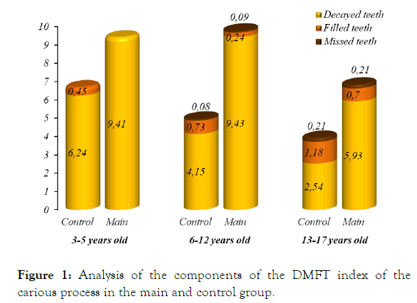
Analysis of the components of the DMFT index showed that the mean number of dental caries lesions in patients with β- thalassemia major was 2.5 times more than in somatically healthy individuals. When studying the degree of caries activity of primary teeth in the first age group in individuals with β- thalassemia major, the degree of caries incidence was 9.41 ± 0.62. In this group, the largest percentage of untreated carious teeth was also noted, which indicates the absence of visits to the dentist at this age. In the control group of the same age, the index of the filled teeth was 0.45 ± 0.70.
An analysis of the indices of caries intensity in the second group indicated that every patient with thalassemia has permanent teeth affected by caries. The total value of the DMFT+DFT was equal to 9.75 ± 0.35 in the main and 4.84 ± 0.25 in the control Component “D” in patients with β-thalassemia major was 2 times higher than this indicator of the control group (p<0.001). The proportion of filled teeth was 0.24 ± 0.05 in the group with β-thalassemia major and 0.76 ± 0.09 in the control group. In the third main group with permanent dentition, the mean value of teeth affected by caries was 5.93 ± 0.24 while the mean value of carious teeth in the healthy group was 2.54 ± 0.2 (p<0.001). The proportion of treated teeth in the group with β- thalassemia major was 33.3 ± 6.2% and in the control group 56.5 ± 5.4%. The status of premature loss of permanent teeth was very low in this study group do not have a statistically significant difference. Prevalence of missing teeth due caries was 0.21 ± 0.06 in both the main and control groups (Figure 1).

Figure 1. Analysis of the components of the DMFT index of the carious process in the main and control group.
Analysis of the components of the DMFT index showed a predominance of the percentage of carious teeth over the filled teeth in the first and second age groups of healthy patients and a significant predominance of untreated teeth in all three groups among patients with β-thalassemia major. Of the 256 adult children, 30 had extracted permanent teeth, which corresponds to 10%. Thus, we found that in children with β-thalassemia major the prevalence and caries intensity of primary teeth is 2 times higher than in practically healthy children. At the same time, the mean number of decayed permanent teeth in thalassemia patients is almost 3 times higher than the mean number of decayed permanent teeth in children of the control group.
Findings and Conclusion
In children with β-thalassemia major, there are high rates of prevalence and intensity of dental caries with a significant predominance of untreated teeth, which indicates the shortcomings in the organization of dental care for such patients. To improve dental care for children with β- thalassemia major, it is necessary to carry out a complex of therapeutic and prophylactic measures, taking into account the age of children, the state of immunity, and the oral biocenosis. It is necessary to conduct parental health education on the limitation of sugar in the diet of children, adherence to diet and thorough, regular tooth brushing with fluoride toothpaste, as well as the need for regular visits to the dentist (at least 3 times a year). It is advisable to organize the work of the dental office in the thalassemia treatment centers with a pre-trained dentist to optimize not only preventive and therapeutic processes but also dispensary observation (group 3) for this category of patients.
REFERENCES
- Weatherall DJ. Keynote address: The challenge of thalassemia for the developing countries. An New York Acad of Sci. 2005;1054(1):11-17.
- Rivella S. Iron metabolism under conditions of ineffective erythropoiesis in β-thalassemia. Blood. 2019;133(1):51-58.
- Orino K, Lehman L, Tsuji Y, Ayaki H, Torti SV, Torti FM. Ferritin and the response to oxidative stress. Biochem J. 2001;357:241-247.
- Bretz WA, Corby P, Schork N, Hart TC. Evidence of a contribution of genetic factors to dental caries risk. J Evid Based Dent Pract. 2003;3(4):185-189.
- Wang X, Willing MC, Marazita ML. Genetic and environmental factors associated with dental caries in children: The Iowa Fluoride Study Caries Research. Karger. 2012;46:3177-184.
- Laurence B, Reid BC, Katz RV. Sickle cell anemia and dental caries: a literature review and pilot study. Spec Care Dentist. 2002;22:70-74.
- Singh J, Singh N, Kumar A, Kedia NB, Agarwal A. Dental and periodontal health status of beta-thalassemia major and sickle cell anemic patients: a comparative study. J Int Oral Health. 2013;5:53-58.
- Al-Wahadni AM, Taani DS, Al-Omari MA. Dental diseases in subjects with beta-thalassemia major. Com Dentist Oral Epidemiol. 2002;30:418-422.
- Hadlinskaya RV, Guliev MR, Gamidova GE. Metabolic aspects with consistent pathology in patients with β-thalassemia major. Siber Med Rev. 2018;6:43-47.
- Cutando SA, Gil Montoya AJ, López-González GJ. Thalassemias and their dental implications. Medicina oral : órgano oficial de la Sociedad Española de Medicina Oral y de la Academia Iberoamericana de Patología y Medicina Bucal. Med Oral. 2002;7:36-40.
- Lugliè PF, Campus G, Deiola C, Mela MG, Gallisai D. Oral condition, chemistry of saliva, and salivary levels of Streptococcus mutans in thalassemic patients. Clin Oral Invest. 2002;6:223-224.
- Aliyeva RQ, Zeynalova GK. Organization of the program for prevention dental diseases in Azerbajan. Int Dental J. 2014;65:92-102.
- Akarslan ZZ, Sadik B, Sadik E. Dietary habits and oral health-related behaviors in relation to DMFT indexes of a group of young adult patients attending a dental school. Med Oral Patol Oral Cir Bucal. 2008;1:13-17.
Citation: Shadlinskaya RV, Zeynalova GK (2019) The Evaluation of the Prevalence and Intensity of Dental Caries in β-thalassemia Major Patients. Dentistry 9:545. doi: 10.35248/2161-1122.19.9.545
Copyright: © 2019 Shadlinskaya RV, et al. This is an open-access article distributed under the terms of the Creative Commons Attribution License, which permits unrestricted use, distribution, and reproduction in any medium, provided the original author and source are credited

