Indexed In
- Open J Gate
- JournalTOCs
- RefSeek
- Hamdard University
- EBSCO A-Z
- OCLC- WorldCat
- Scholarsteer
- Publons
- Geneva Foundation for Medical Education and Research
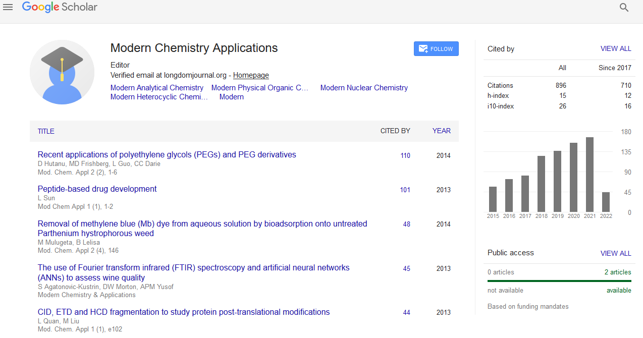
- Google Scholar
Useful Links
Share This Page
Journal Flyer

Open Access Journals
- Agri and Aquaculture
- Biochemistry
- Bioinformatics & Systems Biology
- Business & Management
- Chemistry
- Clinical Sciences
- Engineering
- Food & Nutrition
- General Science
- Genetics & Molecular Biology
- Immunology & Microbiology
- Medical Sciences
- Neuroscience & Psychology
- Nursing & Health Care
- Pharmaceutical Sciences
Perspective Article - (2022) Volume 10, Issue 6
Spectroscopy of Cluster Compounds in DNA and Gene Expression
Yen Caserta*Received: 23-May-2022, Manuscript No. MCA-22-17289; Editor assigned: 26-May-2022, Pre QC No. MCA-22-17289(PQ); Reviewed: 17-Jun-2022, QC No. MCA-22-17289; Revised: 23-Jun-2022, Manuscript No. MCA-22-17289(R); Published: 30-Jun-2022, DOI: 10.35248/2329-6798.22.10.358
Description
Raman spectroscopy has been an active role in the studies of Fe– S proteins of diverse structures and functions. It has been playing an exceptionally active role in the studies of Fe–S proteins of diverse structures and functions. When the wavelength of the excitation laser coincides with that of an allowed electronic transition of the protein chromophore, the intensities of certain Raman bands become selectively enhanced by several orders of magnitude.
Resonance Raman (RR) Spectroscopy has proved to be an indispensable tool for identification and characterization of Fe–S clusters in proteins and in particular those that are diamagnetic. When the molecule is excited with a strong monochromatic light the energy of an electric-dipole moment allows the electronic transition, of a vibronic coupling with the electronically excited state increases the probability.
Fe- S cluster compounds
In DNA t he non-hemic metalloproteins, the enhanced modes mainly contain metal–ligand and intra-ligand stretching and bending vibrations, which include amino acid residues or small inorganic ligands. Resonance Raman (RR) spectra of Fe–S cluster containing proteins, obtained with a laser of wavelength that matches the energy charge transition selectively enhances modes involving the metal–ligand stretching coordinates, which can be observed in the low-frequency about 200–450 cm−1 region. Depending on the type and source, hydrogenases can carry a variable number of different Fe–S clusters that are either essential for the ET and with the binuclear center, constitute the active site. Fe–S cluster biosynthesis has been an active field of research in the last two decades, and RR spectroscopy, in combination with other spectroscopic techniques in several steps of this complex process, which are remarkably conserved in prokaryotic and eukaryotic organisms. There is a growing evidence for the presence of 4Fe–4S clusters in enzymes that take part in nucleic acid processing machinery. These include DNA repair enzymes, such as damage-specific Deoxy Ribose Nucleic Acid (DNA) glycosylases: endonuclease III and their homologue enzymes found in many organisms including humans, as well as primases, helicases, transcription factors, polymerases and Ribose Nucleic Acid (RNA) methyltransferases.
In gene expression the Fe–S clusters are present in transcriptional and translational regulators of gene expression, in which upon environmental stimuli, they undergo transformation that triggers respective cellular response mechanisms. In combination with other spectroscopic techniques, RR has revealed the details about the cluster formation, type and coordination along these processes. These studied systems include proteins that are stressresponsive transcriptional regulators, and/or participate in iron metabolism, such as BolA proteins and homologues. Among all of them Fra2, plays a key role in regulating the iron homeostasis in yeasts. Resonance Raman (RR) spectroscopy revealed the molecular details about the complex formation between Fra2 and cytosolic monothiol glutaredoxins via bridging though cluster. The electronic transitions of Fe–S proteins in ultra and visible region are relatively weak when multiple clusters are present; they result in a broad, featureless spectrum that hinders the assignment of individual clusters. The study of protein has other chromophores, namely heme centers, which have molar extinction coefficients in the visible region approximately ten times higher than Fe–S centers.
Conclusion
Resonance Raman (RR) studies of heme proteins, cobalamin, chlorophylls, carotenoids, flavin nucleotides, the visual pigments DNA, gene expression and bacteriorhodopsin, and a variety of and copper metalloprotein sites have been carried out in laboratories. Spectroscopic techniques, allow us to identify and characterize Fe–S clusters in exceptionally complex, unstable and transient systems and processes. Due to these advances, that have been able to encounter previously undetected Fe-S clusters in known proteins, to propose new roles for the clusters and to improve our understanding of physiological relevant and unusall y complex cofactors that integrate Fe-S clusters.
Citation: Caserta Y (2022) Spectroscopy of Cluster Compounds in DNA and Gene Expression. Modern Chem Appl.10:358.
Copyright: © 2022 Caserta Y. This is an open-access article distributed under the terms of the Creative Commons Attribution License, which permits unrestricted use, distribution, and reproduction in any medium, provided the original author and source are credited.


