Citations : 2345
Dentistry received 2345 citations as per Google Scholar report
Indexed In
- Genamics JournalSeek
- JournalTOCs
- CiteFactor
- Ulrich's Periodicals Directory
- RefSeek
- Hamdard University
- EBSCO A-Z
- Directory of Abstract Indexing for Journals
- OCLC- WorldCat
- Publons
- Geneva Foundation for Medical Education and Research
- Euro Pub
- Google Scholar
Useful Links
Share This Page
Journal Flyer

Open Access Journals
- Agri and Aquaculture
- Biochemistry
- Bioinformatics & Systems Biology
- Business & Management
- Chemistry
- Clinical Sciences
- Engineering
- Food & Nutrition
- General Science
- Genetics & Molecular Biology
- Immunology & Microbiology
- Medical Sciences
- Neuroscience & Psychology
- Nursing & Health Care
- Pharmaceutical Sciences
Review Article - (2024) Volume 14, Issue 3
Smile Esthetics in Orthodontics-Review of Literature
Jibin Joy Daniel* and Anil kumarReceived: 05-Oct-2020, Manuscript No. DCR-24-6711; Editor assigned: 08-Oct-2020, Pre QC No. DCR-24-6711 (PQ); Reviewed: 22-Oct-2020, QC No. DCR-24-6711; Revised: 16-Aug-2024, Manuscript No. DCR-24-6711 (R); Published: 13-Sep-2024, DOI: 10.35248/2161-1122.24.14.701
Abstract
The goal of modern orthodontics is to establish the best possible occlusal relationship between the maxillary and mandibular dentition while maintaining or enhancing facial esthetics. Patient interest in improving facial esthetics is, in part, responsible for the increased growth of the orthodontic practice. One of the most important facial features in predicting attractiveness is smile. As the field and available technologies have continued to evolve, a gradual shift towards an increased emphasis on dental esthetics in treatment planning has occurred and now an esthetically pleasing smile is a key desired outcome of orthodontic treatment. This review article emphysis on components of macro, micro esthetics components in orthodontics.
Keywords
Smile; Macroesthetics; Microesthetics; Orthodontics
Introduction
Beauty is in the mind of the beholder; each mind perceives a different beauty” famously said by writer Margeret Wolfe Hungerford. A beautiful smile is a gateway to the world [1]. The search for improved dentofacial esthetics persists in modern society webster defines the smile as “a change of facial expression involving a brightening of the eyes, an upward curving of the corners of the mouth with no sound and less muscular distortion of the features than in a laugh that may express amusement, pleasure, tender affection, approval, restrained mirth, irony, derision or any of various other emotions.”
The goal of modern orthodontics is to establish the best possible occlusal relationship between the maxillary and mandibular dentition while maintaining or enhancing facial esthetics [2]. Patient interest in improving facial esthetics is, in part, responsible for the increased growth of the orthodontic practice. Physical attractiveness is important social resource in our culture.
One of the most important facial features in predicting attractiveness is smile. This review article emphasis on components of macro, micro esthetics components in orthodontics [3].
Literature Review
Classification of smile esthetics
Macro-esthetics: Here patient profile, vertical proportion, lip fullness, chin-nasal projections, facial widths are considered.
Mini-esthetics: Here assessments of excessive incisor display on smile, smile symmetry, smile arc etc are considered [4].
Micro-esthetics: This includes assessment of tooth proportions in height and width, gingival shape and contour, connectors and embrasures tooth shade etc.
This review article emphasis on components of macro, micro esthetics components in orthodontics.
Discussion
Macroesthetics
The first step in evaluating facial proportions is to take a good look at the patient, examining him or her for developmental characteristics and a general impression [5].
Vertical division: The face is ideally divided into equal thirds.
• Upper: Tragion to opharic
• Middle: Opharic to subnasion
• Lower: Subnasion to gonion
The lower third of the face is further divided into two unequal parts:
• Subnasion to commissures of the lips is equal to one-third or
18 mm to 20 mm from the subnasion to the upper lip [6].
• Commissures of the lips to the gonion is equal to two-thirds
or 36 mm to 40 mm from the lower lip to the gonion (Figure
1).
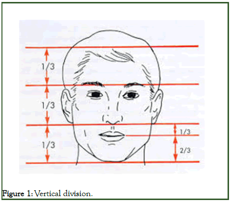
Figure 1: Vertical division.
Vertical facial proportions in the frontal and lateral views are best evaluated in the context of the facial thirds. Changes in lower third of the face.
Increase lower one-third height:
• Vertical maxillary excess
• Class III malocclusion
Decreased lower one-third height:
• Vertical maxillary deficiencies
• Mandibular retrusion bite
Horizontal division
An ideally proportional can be divided into central, medial and lateral equal fifths. The separation of the eyes and the width of the eyes, which should be equal, determine the central and medial fifths [7]. The nose and chin should be centered within the central fifth, with the width of the nose the same as or slightly wider than the central fifth (Figure 2).
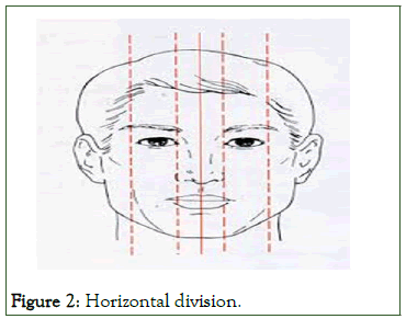
Figure 2: Horizontal division.
Balance and symmetry
Facial symmetry is defined by the facial midline. The midline runs through the center of the face and a philtrum of the lip (cupid’s bow), dividing it into right and left sides. The more symmetric and identical the sides, the closer they come to bilateral duplication or mirror images, the more inherently harmonious and beautiful the face (horizontal symmetry). This is the opposite of the dental midline, which seeks beauty through diversity (radiating symmetry) (Figure 3).
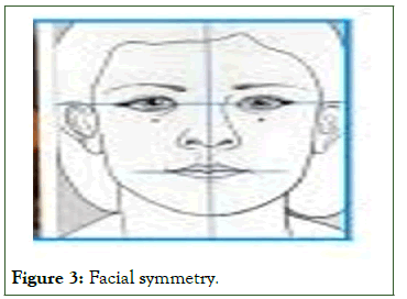
Figure 3: Facial symmetry.
Facial profile
This step requires placing the patient in the physiologic natural head position, the head position the individual adopts in the absence of other cues. This can be done with the patient either sitting upright or standing, but not reclining in a dental chair and looking at the horizon or a distant object. With the head in this position, note the relationship between two lines, one dropped from the bridge of the nose to the base of the upper lip and a second one extending from that point downward to the chin. These line segments should form a nearly straight line. An angle between them indicates either profile convexity (upper jaw prominent relative to chin) or profile concavity (upper jaw behind chin) [8].
A convex profile therefore indicates skeletal class II jaw relationship. A concave profile indicates a skeletal class III jaw relationship. If the profile is approximately straight, it does not matter whether it slopes either anteriorly (anterior divergence) or posteriorly (posterior divergence). Divergence of the face (the term was coined by the eminent orthodontist-anthropologist Milo Hellman) is influenced by the patient's racial and ethnic background (Figure 4).
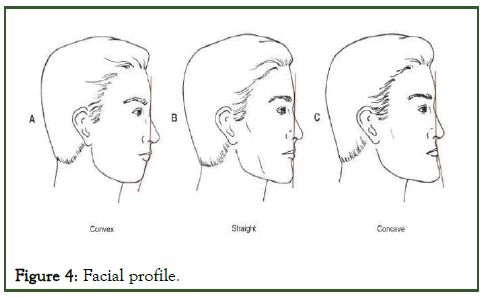
Figure 4: Facial profile.
Microesthetics
Subtleties in the proportions and shape of the teeth and associated gingival contours have been emphasized in the burgeoning literature on "cosmetic dentistry" in recent years. A similar evaluation is necessary in the development of an orthodontic problem list if an optimal esthetic result is to be obtained [9].
Width and height of crowns: There is a variation on dental dimensions that can be considered as normal or desirable, especially when considering that there are individuals with different facial patterns. Some works show that the dimensions of anterior teeth can be defined within a limited range. The height of the central incisor varies from 10.4 mm to 11.2 mm while its width varies from 8.73 mm to 9.3 mm and usually, these references are used in prosthetic reconstructions, when no other parameters are available.
The most important is the correlation of these dimensions, i.e., the dental proportions. Two proportions must be considered: The relation between height and width of each tooth and the relation of height and width among the teeth. In these cases, we will always be referring to the real dimension of the teeth, i.e., the clinical crown dimensions of anterior teeth [10].
A classic study that investigated dental proportions was that by Gillen et al., who found the following proportions of width among the upper anterior teeth: a) lateral incisors have 78% of the width of the central incisor (lateral incisor=central incisor × 0.78); b) lateral incisor has 87% of the width of the canine (lateral incisor=canine × 0.87); c) canine has 90% of the width of the central incisor (canine=central incisor × 0.90).
Most authors define the height/width ratio of 0.80 for the upper central incisor (which represents the key tooth to esthetical composition of the smile) as standard to be used in prosthesis, periodontics and orthodontics. Therefore, on establishing ideal widths, based on intact teeth, it is suggested to use the proportion of 80 ± 5% to define the ideal height, relating it to the facial pattern and to the individual’s natural dental proportions (Figure 5).
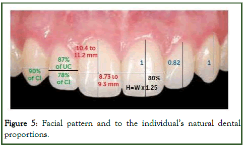
Figure 5: Facial pattern and to the individual’s natural dental proportions.
The relation of proportion between the crown heights of anterior teeth proposed by Gillen et al., is widely used. It suggests that the height of the clinical crown of the upper lateral incisor must be 82% of the height of the crowns of the central incisor and canine. Therefore, canines and upper central incisors would have the same anatomical crown height. This study was used to justify the bonding of orthodontic brackets at the same height for canines and upper central incisors, during placement of the orthodontic appliance [11].
In an attractive smile, the apparent width of the lateral incisor is 62% of the width of central incisor, width of canine is 62% of the width of lateral incisor and1st premolar is 62% of the width of canine. The apparent width of maxillary anterior teeth on smile and their actual mesio-distal width, differ because of the curvature of dental arch. This recurring 62% proportion also appears in other relationship in human anatomy and is known as golden proportions. It is an excellent guideline in day to day practice in finishing cases when lateral incisors are disproportionately small or are missing (Figure 6).
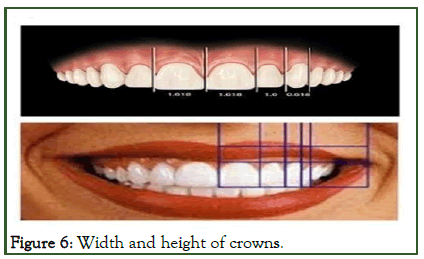
Figure 6: Width and height of crowns.
Connector area and embrasures: Morley and Eubank recently introduced the term connector area as a useful tool and a visual goal to optimize smile esthetics in dental patients. Connector areas are larger, broader areas than the contact points between teeth and can be defined as the zone in which two adjacent teeth appear to touch.
The most esthetic relationship of connector area between the maxillary anterior teeth is referred to as the 50-40-30 rule.
This rule defines the ideal connector area between the two maxillary central incisors as 50% of their clinical crown length, the ideal connector area between central and lateral is 40% of central incisor clinical crown length and between lateral and canine it is 30% of clinical crown length of central incisor (Figure 7).
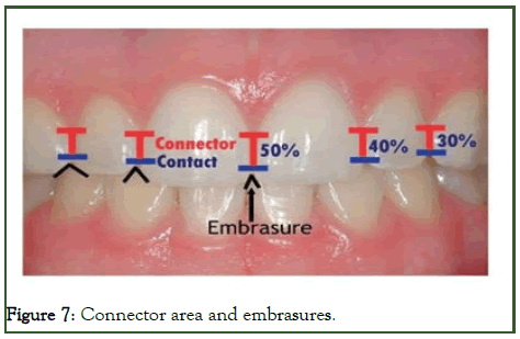
Figure 7: Connector area and embrasures.
The most important connector area is the one between two maxillary central incisors and should be maintained in orthodontically treated cases.
The embrasures (the triangular spaces incisal and gingival to the contact) ideally are larger in size than the connectors and the gingival embrasures are filled by the interdental papillae.
Short interdental papillae leave an open gingival embrasure above the connectors called “black triangles” which give an unesthetic appearance of the teeth during smile.
In adults, they arise due to loss of gingival tissue but when crowded and rotated maxillary incisors are corrected orthodontically, the connectors move incisal and black triangles may appear.
Reshaping of teeth by orthodontic root paralleling and flattening of the mesial surfaces of the central incisors, followed by space closure, will lengthen this contact area and move it apically toward the papilla and correct the black triangles.
Tooth shade and colour: Aging brings about changes in the tooth colour and shade. In young age teeth appear lighter and brighter. As age advances the teeth appear darker and duller. This is due to the formation of secondary dentin as pulp chambers decrease in size and thinning of enamel leads to decrease in its transparency and a greater contribution to darker shade.
A normal progression of shade changes from midline posteriorly is an important contributor to an attractive and natural appearing smiles. The maxillary central incisors tend to be the brightest in a smile, lateral incisor less bright and canines the least, 1st and 2nd premolar are lighter and brighter than the canines and closely match lateral incisors.
Gingival height, shape and contours: Proportional gingival height is necessary to produce a normal and attractive dental appearance. The central incisor has the highest gingival level, lateral incisor is 1.5 mm incisal and canine is at the same level as central incisor. A difference of more than 2 mm in the gingival height is obvious. This is important in finishing all orthodontic cases and also when tooth substitutions are planned. Gingival shape refers to curvature of gingiva at the margin of the tooth (Figure 8) [12].
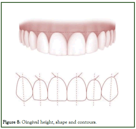
Figure 8: Gingival height, shape and contours.
According to the accreditation criteria for the American academy of cosmetic dentistry “the shape of maxillary lateral incisor and mandibular incisors is symmetrical and half oval. Shape of maxillary centrals and canines are more elliptical and asymmetric”. Gingival zenith is the most apical point of gingival tissue and should be located distal to the longitudinal axis of maxillary centrals and canines while zenith of maxillary lateral and mandibular incisor should coincide with their long axis.
Correcting the gingival contours removes the excess gingival covering the clinical crowns and helps improve display of teeth and improves gingival contours and shape. This is carried out with gingivectomy procedure.
Gingival contouring can now be carried out effectively with the use of a diode lasers.
Conclusion
Today, well occluding casts and pleasing profiles can no longer be considered to be adequate treatment goals for the orthodontist. Since most people interact with each other facing each other directly or obliquely, the smile of the patient should be given adequate importance in treatment planning. In order to correctly diagnose and treat problems associated with the smile, meticulous clinical observation and record taking in the form of photos and videos is warranted, in various dimensions.
In achieving smile aesthetics, dental elements, facial elements, gums related factors and physical elements should be evaluated collectively. Treatments should be applied in a unified frame.
These concepts of smile esthetics are not new, but are too often overlooked in orthodontic treatment planning. The ideal components of the smile should be considered not as rigid boundaries, but as artistic guidelines to help orthodontists treat individual patients who are today, more than ever, highly aware of smile esthetics.
Although there are many things to consider when planning an esthetic case, many principles and guidelines exist that can help direct treatment. It is important to have a good understanding of the overriding ethical principles as well as the elements of macro, mini and micro-esthetics prior to performing esthetic dentistry.
A simple rule of thumb is to start with the large features and work toward the smaller features. Look at the face, lips and gingiva before individual tooth assessments are performed. In other words, look at the forest before you look at the trees. A sound knowledge of orthodontic mechanics and growth changes as well as the principles of cosmetic dentistry should help the orthodontist to truly live up to the epithet of smile architect.
References
- Morley J, Eubank J. Macroesthetic elements of smile design. J Am Dent Assoc. 2001;132(1):39-45.
[Crossref] [Google Scholar] [PubMed]
- Rinchuse DJ, Kandasamy S. Centric relation: A historical and contemporary orthodontic perspective. J Am Dent Assoc (1939). 2006;137(4):494-501.
[Crossref] [Google Scholar] [PubMed]
- Sarver DM. Principles of cosmetic dentistry in orthodontics: Part 1. Shape and proportionality of anterior teeth. Am J Orthod Dentofacial Orthop. 2004;126(6):749-753.
[Crossref] [Google Scholar] [PubMed]
- Sabri R. The eight components of a balanced smile. J Clin Orthod. 2005;39(3):155-154.
[Google Scholar] [PubMed]
- Takenoshita M, Miura A, Shinohara Y, Mikuzuki R, Sugawara S, Tu TT, et al. Clinical features of atypical odontalgia; three cases and literature reviews. Biopsychosoc Med. 2017;11:21.
[Crossref] [Google Scholar] [PubMed]
- Sterrett JD, Oliver T, Robinson F, Fortson W, Knaak B, Russell CM. Width/length ratios of normal clinical crowns of the maxillary anterior dentition in man. J Clin Periodontol. 1999;26(3):153-157.
[Crossref] [Google Scholar] [PubMed]
- Sarver DM, Yanosky M. Principles of cosmetic dentistry in orthodontics: Part 2. Soft tissue laser technology and cosmetic gingival contouring. Am J Orthod Dentofacial Orthop. 2005;127(1):85-90.
[Crossref] [Google Scholar] [PubMed]
- Beyer JW, Lindauer SJ. Evaluation of dental midline position. Semin Orthod. 1998;4(1):146-152.
[Crossref] [Google Scholar] [PubMed]
- Pini NP, de-Marchi LM, Gribel BF, Ubaldini AL, Pascotto RC. Analysis of the golden proportion and width/height ratios of maxillary anterior dentition in patients with lateral incisor agenesis. J Esthet Restor Dent. 2012;24(6):402-414.
[Crossref] [Google Scholar] [PubMed]
- Chu SJ. Range and mean distribution frequency of individual tooth width of the maxillary anterior dentition. Pract Proced Aesthet Dent. 2007;19(4):209-215.
[Google Scholar] [PubMed]
- Godinho J, Gonçalves RP, Jardim L. Contribution of facial components to the attractiveness of the smiling face in male and female patients: A cross-sectional correlation study. Am J Orthod Dentofacial Orthop. 2020;157(1):98-104.
[Crossref] [Google Scholar] [PubMed]
- Benoliel R, Gaul C. Persistent idiopathic facial pain. Cephalalgia. 2017;37(7):680-691.
[Crossref] [Google Scholar] [PubMed]
Citation: Daniel JJ, Kumar A (2024) Smile Esthetics in Orthodontics- Review of Literature. J Dentistry. 14:701.
Copyright: © 2024 Daniel JJ, et al. This is an open-access article distributed under the terms of the Creative Commons Attribution License, which permits unrestricted use, distribution and reproduction in any medium, provided the original author and source are credited.
