Indexed In
- Open J Gate
- Genamics JournalSeek
- JournalTOCs
- Ulrich's Periodicals Directory
- RefSeek
- Hamdard University
- EBSCO A-Z
- OCLC- WorldCat
- Publons
- Geneva Foundation for Medical Education and Research
- Euro Pub
- Google Scholar
Useful Links
Share This Page
Journal Flyer

Open Access Journals
- Agri and Aquaculture
- Biochemistry
- Bioinformatics & Systems Biology
- Business & Management
- Chemistry
- Clinical Sciences
- Engineering
- Food & Nutrition
- General Science
- Genetics & Molecular Biology
- Immunology & Microbiology
- Medical Sciences
- Neuroscience & Psychology
- Nursing & Health Care
- Pharmaceutical Sciences
Case Report - (2024) Volume 15, Issue 2
Primary Malignant Neoplasm of Ectopic Thyroid Tissue: A Rare Pathology
Xavier Garnica*, Angel Borges and Jose Felix VivasReceived: 05-Jan-2024, Manuscript No. JCM-24-24660; Editor assigned: 08-Jan-2024, Pre QC No. JCM-24-24660 (PQ); Reviewed: 22-Jan-2024, QC No. JCM-24-24660; Revised: 29-Jan-2024, Manuscript No. JCM-24-24660 (R); Published: 05-Feb-2024, DOI: 10.35248/2157-2518.24.15.440
Abstract
The presence of thyroid ectopic tissue arises from an alteration in the embryological development of said gland, and may have aberrant locations throughout the human economy. The possibility of developing cancer in that tissue is highly infrequent, however, it is described in the literature. We present the case of a female patient who debuted with respiratory symptoms in relation to an occupying lesion in the middle mediastinum and the presence of cervical adenomegaly, from which a biopsy was taken and it was positive for metastatic papillary carcinoma of the thyroid, however, in preoperative studies the thyroid was found without alterations; then he presented a tumor in the sacral region whose histology is consistent with that mentioned above, which is why a total thyroidectomy was planned, the surgical specimen being negative for malignancy. In the presence of an unrespectable papillary carcinoma of ectopic thyroid tissue located in the mediastinum, the patient is referred to nuclear medicine to begin treatment with radioactive iodine and subsequently evaluate response.
Keywords
Papillary carcinoma; ectopic thyroid tissue; thyroidectomy
Introduction
Ectopic thyroid tissue is the most common type of dysgenesis that occurs in relation to the thyroid gland and is usually found along the vestige of the obliterated thyroglossal duct, usually from the base of the tongue to the mediastinum, however, in the literature there are numerous reports of quite particular foreign locations [1]. Its risk of malignancy is less than 1%, making it a truly rare nosological entity, which is why this case is brought up.
Case Presentation
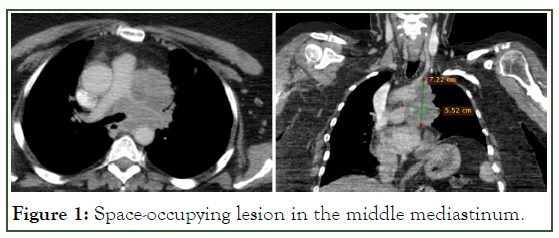
A 52-year-old female patient who began her current illness in April 2020 when she presented with intermittent heartburn associated with postprandial fullness, dry non-productive cough, and progressive dysphonia, which is why she went to a doctor for evaluation. Non-contributory personal history. Physical examination is within normal limits. They performed nasofibrolaryngoscopy and reported paramedian paralysis of the left vocal cord as a finding; Later, a tomographic study showed evidence of a 7 × 5cm space-occupying lesion in the middle mediastinum in close relationship with the great thoracic vessels and generating extrinsic occlusion of the left pulmonary artery and the left main bronchus in 80% (Figure 1).

Figure 1: Space-occupying lesion in the middle mediastinum.
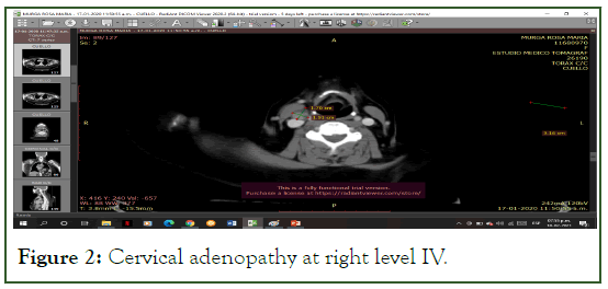
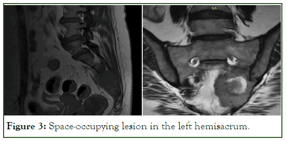
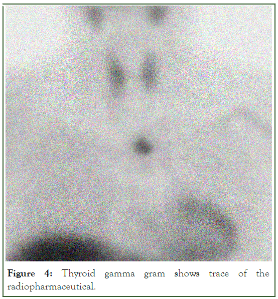
Visualized lymphadenopathy at right cervical level IV (Figure 2), which is why a lateral cervicotomy was performed with an excisional biopsy, showing positivity for metastatic papillary carcinoma of thyroid origin according to an immunohistochemical study (corroborated twice). Thyroid ultrasound and thyroid tests without abnormalities. Elevated thyroglobulin (35,600 ng/mL). In view of presenting respiratory failure of abrupt onset, it was decided to perform 3 doses of emergency radiotherapy, improving symptoms. Subsequently, he presented pain and altered sensitivity in the left lower limb, an MRI-type study of the lumbosacral spine was performed and a space-occupying lesion of 8 × 7cm was evident in the left sacrum (Figure 3), oppressing ipsilateral nerve roots, a left hemisacrectomy was performed and the result of the biopsy was performed was positive for poorly differentiated metastatic papillary carcinoma. Oncodeep® was performed on lymph node and sacral tumor and both were positive for papillary neoplasia of thyroid origin. Thyroid scan shows trace of the radiopharmaceutical in the usual location and at the mediastinal level (Figure 4). Total thyroidectomy was planned and was performed without complications, the biopsy of which was negative for malignancy.

Figure 2: Cervical adenopathy at right level IV.

Figure 3: Space-occupying lesion in the left hemisacrum.

Figure 4: Thyroid gamma gram shows trace of the radiopharmaceutical.
It was decided to refer to the nuclear medicine service to begin ablative therapy with radioactive iodine given that the mediastinal lesion does not have respectability criteria and subsequently evaluate response to treatment.
Discussion
It is estimated that the prevalence of ectopic thyroid tissue is 1 per 300,000 people, with a predilection for the female sex (75%) as in most thyroid pathologies [1]. They usually present asymptomatic, however, the clinical manifestations when they occur are associated with the size and location of the lesion. Primary carcinomas arising from ectopic thyroid tissue are rare and have been reported to arise from thyroid tissue in thyroglossal cysts, aberrant cervical thyroid tissue, lingual thyroid, mediastinum, and struma ovarii [2]. Regarding the intrathoracic location, Nguyen, et al. 3 reported a case of a 90-year-old male patient with anaplastic carcinoma of ectopic thyroid tissue at the mediastinal level, which presented as a lesion that invaded the sternum and the overlying skin, noting the aggressive nature of this histological subtype especially in the male gender [3].
Differentiating between carcinoma arising in ectopic thyroid tissue and metastatic carcinoma is difficult; the diagnosis can be made indirectly taking into account some characteristics such as a separate blood supply to the ectopic gland, no personal history of malignancy, and a normal or absent orthotopic thyroid without a history of surgery [2]. On many occasions, the resection of an extra thyroidal lesion is performed and the biopsy is positive for neoplasia of thyroid origin; in this case, the orthotopic thyroid tissue should be investigated if there is any suspicion of malignancy, the most appropriate way being, after the imaging studies, the histopathological analysis of the definitive specimen after a total thyroidectomy. If there is no report of malignancy in the biopsy, we are faced with the presence of a neoplasm of ectopic thyroid tissue, as described by Qi et al [4]. In their case report of a 50-year-old female patient with two lung nodules of metastatic appearance, which after excision showed a thyroid carcinoma, so they decided to perform a total thyroidectomy, not showing any malignancy in the definitive specimen. Another tool to differentiate between metastasis and ectopic thyroid neoplasia is with certain immunohistochemical markers such as CK19, Gal-3 and HBME-1 (the latter being the most specific), which are positive in the extra thyroid neoplastic areas but negative in the tissue Intrathyroid [5].
The standard for the diagnosis of these pathologies is the thyroid scan, which is highly specified to demonstrate the presence of thyroid tissue in erratic locations [6]. Failing this, positron emission tomography is also a diagnostic option that allows visualization of abnormal uptake of ectopic thyroid tissue [7].
Regarding treatment, surgical resection is the preferred modality for both orthotopic and ectopic thyroid tissue, with subsequent radioactive iodine therapy if warranted [2,6]. If the ectopic neoplasm is located near the thyroid gland, en bloc resection of both elements can be performed, such as a sistrunk procedure +total thyroidectomy [8]. In the case where the lesion has unresectable criteria or the patient is medically inoperable due to some medical condition or simply refuses the surgical procedure, radioactive iodine therapy (I131) would also be a valid treatment option [9]. In the literature, many cases of occult papillary carcinomas are described that clinically debut with distant metastasis; however, cases where the orthotropic thyroid gland is free of malignancy are extremely rare, the primary being purely ectopic tissue [10].
Conclusion
Primary carcinoma of ectopic thyroid tissue is an extremely rare oncological entity which warrants high clinical suspicion. The thyroid scan represents the main diagnostic method in these cases, however, it does not allow you to discern its biological behavior. Surgical resection is the main therapeutic option and subsequently, if necessary, ablative therapy with radioactive iodine.
References
- Santangelo G, Pellino G, De Falco N, Colella G, Amato DS, Maglione MG, et al. Prevalence, diagnosis and management of ectopic thyroid glands. Int J Surg. 2016;28:S1-6.
[Crossref] [Google Scholar] [PubMed]
- Karim F, Inam H, Choudry UK, Hasnain Fatimi S. Ectopic follicular variant of papillary thyroid carcinoma in anterior mediastinum with a normal thyroid gland. A case report. Int J Surg Case Rep. 2018;51:213-217.
[Crossref] [Google Scholar] [PubMed]
- Nguyen D, Htun NN, Wang B, Lee B, Johnson C. An anaplastic thyroid carcinoma of the giant-cell type from a mediastinal ectopic thyroid gland. Diagnostics. 2023;13:2941.
[Crossref] [Google Scholar] [PubMed]
- Qi Y, Liu J, Liu Y, Shen Z, Hu N. Ectopic papillary thyroid carcinoma mimicking distant metastatic tissue. J Int Med Res. 2022;50(9):1-6.
[Crossref] [Google Scholar] [PubMed]
- Cabibi D, Cacciatore M, Guarnotta C, Aragona F. Immunohistochemistry differentiates papillary thyroid carcinoma arising in ectopic thyroid tissue from secondary lymph node metastases. Thyroid. 2007;17(7):603-607.
[Crossref] [Google Scholar] [PubMed]
- Shafiee S, Sadrizade A, Jafarian A, Zakavi SR, Ayati N. Ectopic papillary thyroid carcinoma in the mediastinum without any tumoral involvement in the thyroid gland A Case report. Asia Oceania J Nucl Med Biol. 2013;1(1):44-46.
[Crossref] [Google Scholar] [PubMed]
- Huang NS, Wei WJ, Qu N, Wang YL, Wang Y, Ji QH. Lingual ectopic papillary thyroid carcinoma: two case reports and review of the literature. Oral Oncol. 2019;88:186-189.
[Crossref] [Google Scholar] [PubMed]
- Nambiar G, Eshwarappa H, Kini H, Chidanand D. Isolated thyroid carcinoma in an ectopic thyroid tissue. BMJ Case Rep. 2021;14(2):1-4.
[Crossref] [Google Scholar] [PubMed]
- Sturniolo G, Violi MA, Galletti B, Baldari S, Campennì A, Vermiglio F, et al. Differentiated thyroid carcinoma in lingual thyroid. Endocrine. 2016;51(1):189-98.
[Crossref] [Google Scholar] [PubMed]
- Toda S, Iwasaki H, Suganuma N, Okubo Y, Hayashi H, Masudo K, et al. Occult thyroid carcinoma without malignant thyroid gland findings during preoperative examination: report of three cases. Case Rep Endocrinol. 2020;1-6.
[Crossref] [Google Scholar] [PubMed]
Citation: Garnica X (2024) Primary Malignant Neoplasm of Ectopic Thyroid Tissue: A rare pathology. J Carcinog Mutagen. 15:440.
Copyright: © 2024 Garnica X. This is an open-access article distributed under the terms of the Creative Commons Attribution License, which permits unrestricted use, distribution, and reproduction in any medium, provided the original author and source are credited.


