Indexed In
- Academic Journals Database
- Open J Gate
- Genamics JournalSeek
- JournalTOCs
- China National Knowledge Infrastructure (CNKI)
- Scimago
- Ulrich's Periodicals Directory
- RefSeek
- Hamdard University
- EBSCO A-Z
- OCLC- WorldCat
- Publons
- MIAR
- University Grants Commission
- Geneva Foundation for Medical Education and Research
- Euro Pub
- Google Scholar
Useful Links
Share This Page
Open Access Journals
- Agri and Aquaculture
- Biochemistry
- Bioinformatics & Systems Biology
- Business & Management
- Chemistry
- Clinical Sciences
- Engineering
- Food & Nutrition
- General Science
- Genetics & Molecular Biology
- Immunology & Microbiology
- Medical Sciences
- Neuroscience & Psychology
- Nursing & Health Care
- Pharmaceutical Sciences
Research Article - (2024) Volume 15, Issue 2
Potentiation of Foot and Mouth Disease Vaccine by Irradiating Mid-Infrared Rays-A Laser Adjuvant Technology
Uma Kanthan T1*, Madhu Mathi2, Uma Devi3 and Siva Ramakrishnan42Department of Veterinary Medicine, Veterinary Hospital, Vadakupudhu Palayam Post, Erode, Tamil Nadu, India
3Department of Botany, The Standard Fireworks Rajaratnam College for Women, Tamil Nadu, India
4Veterinary Assistant Surgeon, Veterinary Dispensary, Puliyal Post, Devakottai Taluk, Sivaganga, Tamil Nadu, India
Received: 13-Feb-2024, Manuscript No. JVV-24-24884; Editor assigned: 15-Feb-2024, Pre QC No. JVV-24-24884 (PQ); Reviewed: 01-Mar-2024, QC No. JVV-24-24884; Revised: 08-Mar-2024, Manuscript No. JVV-24-24884 (R); Published: 15-Mar-2024, DOI: 10.35248/2157-7560.24.15.558
Abstract
Vaccination is a safe method of disease prevention. Adjuvants that are used in vaccines enhance the immune response against vaccines at a certain level. The prevalence of using infrared in therapeutics is growing, and this necessitates the chance of employing it in vaccine technology to improve the vaccine potency and efficacy. In this study, we invented a 2-6 μm mid-Infrared (mid-IR) ray-generating atomizer, named MIRGA, and applied it to the Foot and Mouth Disease (FMD) vaccine only by externally spraying over the vial. The irradiation is found to have enhanced the titer of post-vaccination antibodies by several folds. Through various instrumentations, we demonstrated the photostimulation and photobiomodulation effects in the vaccine constituents caused by the applied mid-IR radiation. This technology is simple; it helps save money and resources, and it does not have any negative side effects. In the future, it could be an additional method or adjuvant for the potentiation vaccines.
Keywords
Antibodies; Enhancement; Safe; Economy
Abbreviations
MIRGA-Mid-Infrared Generating Atomizer; FMD-Foot and Mouth Disease; EMW-Electromagnetic Wave; LC-MS-Liquid Chromatography-Mass Spectrometry; FT-IR-Fourier-Transform Infrared Spectroscopy; NMRNuclear Magnetic Resonance
Introduction
Vaccination prevents and controls infectious diseases. Adjuvants enhance the immunogenicity of the vaccine. Adjuvant-a Latin word, adjuvare: Means support or enhance it is used as a carrier of an antigen. In 1920 Gaston Ramon-a French veterinarian trialed and framed the concept of the adjuvant [1]. Followed, in 1926 alum adjuvants, and 1940 oil emulsion adjuvants were developed. From 1926 to 1990 alum adjuvants were licensed and used and in 1997 oil adjuvants were licensed. In the following 26 years, many adjuvants were developed and licensed [2].
FMD causes economic loss globally [3]. Seven serotypes and many subtypes exist in the FMD virus. The antibodies are mainly typespecific and sometimes subtype-specific. Currently, inactivated vaccines along with adjuvants are used in commercial vaccines. In India, most trivalent vaccines (O, A, Asia 1) are being used. Over the last 90 years, various adjuvants have been tried and few of them are licensed, but many failed. As a laser adjuvant, we applied 2-6 μm mid-IR over the bottles containing an FMD vaccine. The acquired changes in the chemistry of vaccine constituents and benefits are detailed here.
In 2013, [4] used near-infrared light to illuminate the skin to achieve an effective immunological response. Discuss the medical applications of infrared explaining the photostimulation and photobiomodulation effects of IR [5]. Infrared light also potentiates antimicrobial Indium Phosphide Quantum Dots to eliminate multidrug-resistant pathogens [6]. Recently [7], the infrared for activation of cancer cell vaccine. Considering the growing safe use of infrared radiation, we conceived the thought of using it in vaccine potentiation. More specifically mid-IR is a green field wavelength range.
In the Electromagnetic Wave (EMW) spectrum, the mid-IR region is vital and interesting for many applications since that region coincides with the internal vibration of most molecules [8]. Almost all thermal radiation on the Earth’s surface lies in the mid-IR region, 66% of the sun’s energy we receive is infrared [9] which is absorbed and radiated by all particles on the Earth. Naturally, at the molecular level, the interaction of mid-IR wavelength energy elicits rotational and vibrational modes (from about 4500- 5000cm-1 roughly 2.2 to 20 microns) through a change in dipole movement leading to chemical bond alteration [10]. Mid-infrared is a biologically safe range in the electromagnetic spectrum and coincides with the frequency of all biomolecules [11-13]. The mid- IR can penetrate a variety of natural/anthropogenic obscurants [14], and is being used to treat various diseases in humans [15] and veterinary [16] medicine fields. Considering these applications, we designed an atomizer to generate mid-IR by spraying an imbalanced ionic solution at a constant pressure. FMD vaccine is subjected to the MIRGA spraying and the generated mid-IR alters the molecular configurations resulting in enhanced efficacy of the vaccine.
Materials and Methods
Mid-IR Generating Atomizer (MIRGA) (patent no: 401387) is a 20 ml pocket-sized atomizer (Supplementary file-Figure S1) containing inorganic water-based solution in which approximately two sextillion cations and three sextillion anions are contained; every time spraying emits 0.06 ml which contains approximately seven quintillion cations and eleven quintillion anions. During spraying, depending on the pressure (varies with the user) applied to the plunger, every spraying generates 2-6 μm mid-IR. The features of MIRGA (patent granted 401387) and the detailed process of fabricating it to generate 2-6 μm mid-IR while spraying is discussed by [17-19] (details presented in supplementary Text T1) (raw data of estimation of the generated 2-6 μm mid-IR available in Supplementary data D1).
The mid-IR generated by the inorganic compounds has potential applications in biomedicine [20,21]. Also, it is a new synthesis technique to produce functional materials (2-6 μm mid-IR) [22,23]. It is well-known that combining various compounds with excellent electrical properties creates new composite materials, which have garnered a lot of attention from the technology community recently [24,25]. Trivalent FMD vaccine vials available in India, all with the same brand and batch number, were used in 30 crossbred cattle. The instruments used in this research to identify the following:
Chemical compound transformation: Liquid Chromatography- Mass Spectrometry (LC-MS): Mass spectra were recorded on BrukermicroTOF-Q II Spectrometer. Around 500 microliters of methanol were added to 500 microliters of vaccine and were mixed properly. The solution was then filtered using the 0.22 μM syringe filter (Abdos). Around 40 μl of the filtered sample was injected into the ESI column and the data was recorded.
Chemical bond changes: Fourier-Transform Infrared Spectroscopy (FT-IR): IR spectra were recorded in Thermo scientific iDi transmission accessory, Nicolet IS5 with the software OMNIC. The resolution of the spectrum was recorded with resolution 2 and the number of scans was 16. The scan was done in the wavenumber range of 400 to 4000 cm-1 with the data spacing of 0.241 cm-1. The detector of the instrument was DTGS KBr and the vaccine sample was placed in the IR beam with the liquid sample holder.
Proton resonances: Proton Nuclear Magnetic Resonance (1H-NMR): 1H, spectra were recorded at Bruker AV-400 (1H: 400 MHz, probe-ppb, shim coil temperature 299, sample (vaccine) temperature 25oC, software TOPSPIN). 1H NMR chemical shifts were reported in ppm downfield from tetramethylsilane and 1H NMR spectra were recorded in 10% D2O and were referenced to signals of deuteron solvents. Multiplicity was abbreviated as s-Singlet; d-doublet; dd-doublet of doublets; dt-doublet of triplets; t-triplet; q-quartet; dq, doublet of quartets; td-triplet of doublets; m-multiplet; br-broad.
The MIRGA solution was sprayed from 0.25-0.50 meter towards the packaged (polythene) material (vaccine) (Supplementary video V1). This distance is essential for the MIRGA sprayed solution to form ion clouds, oscillation, and mid-IR generation. The generated 2-6 μm mid-IR could penetrate the intervening polythene package and act on the inside vaccine. Close spraying never generates energy. MIRGA can be used like a body spray.
Thirty cattle (24 vaccinated and 6 control cattle) of both sexes aged between 4 to 5 years were randomly selected from a private farm. The 30 cattle were divided into five groups (A, B, C, D, and E), with 6 cattle in each A, B, C, and D; and 6 in control group E.
The vaccine used in this study was polyvalent FMD. Five vials of the vaccine were divided into five groups with one vial for each group marked separately (i.e., 1, 2, 3, 4, and C). The vial in group 1 was given 1-time MIRGA spraying from a distance of 0.25-0.50 meter towards the vaccine vial; following this, 2, 3, and 4 MIRGA spraying were given to the vials in groups 2, 3, and 4 respectively. The vial in group C received no spraying and served as a control.
We employed multiple sprayings up to 4 times because, in nature, the input of extra energy should always denature the receptor (vaccine).
Cattle in groups A, B, C, and D were inoculated with vaccines that had been sprayed 1, 2, 3, and 4 times, respectively. The control cattle (group E) were inoculated with the vaccine without MIRGA treatment (group C vial). On the 0-day and 21st-day post-vaccination, serum samples were collected from all the animals and analyzed for anti-FMDV antibodies by Liquid-Phase Blocking ELISA (LPBE) [26].
Results and Discussion
The results are expressed in Table 1. Between the animals of each group, the difference in antibody titer was the least, hence the average given (Table 1). To summarize, compared with the control, enhancement of post-vaccination antibody titer could be seen in animals vaccinated with 1, 2, and 3 sprayings, but not in the 4 sprayed vaccine groups. Among these groups, animals vaccinated with 2 sprayings showed the highest antibody titers. (Table 1) (Supplementary Table 1)
| Group | Type O | Type A | Type Asia 1 | |||||||||
|---|---|---|---|---|---|---|---|---|---|---|---|---|
| Pre Vac titre (log 10) | Post Vac titre (log 10) | Change in titer (Log Difference) |
Fold change | Pre Vac titre (log 10) | Post Vac titre (log 10) | Change in titer (Log Difference) |
Fold change | Pre Vac titre (log 10) | Post Vac titre (log 10) | Change in titer (Log Difference) |
Fold change | |
| I | 1.16 | 2.02 | 0.85 | 7.7 | 0.84 | 1.76 | 0.91 | 12.42 | 1.64 | 2.34 | 0.7 | 5.16 |
| II | 1 | 2.17 | 1.89 | 16.56 | 0.73 | 2.08 | 1.35 | 28.41 | 1 | 2.52 | 1.52 | 33.18 |
| III | 0.84 | 1.67 | 1.12 | 7.68 | 0.84 | 1.49 | 0.68 | 6.96 | 1.18 | 2.11 | 0.91 | 8.75 |
| IV | 1.22 | 0.67 | -0.55 | 0.35 | 1 | 1 | 0 | 1 | 1 | 1.36 | 0.13 | 1.41 |
| V (control) |
2.02 | 2.6 | 0.58 | 3.8 | 1.87 | 2.22 | 0.35 | 2.24 | 2.31 | 2.5 | 0.19 | 1.55 |
Table 1: Anti-FMDV antibodies in cattle vaccinated with trivalent vaccine treated with MIRGA.
Instrumentation results
Since LC-MS, FTIR, and NMR observations of 1 sprayed sample are very similar to that of the control, only the control’s results are narrated.
LC-MS
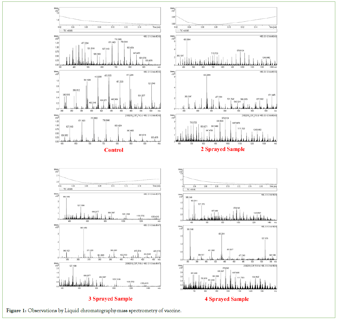
The peak at m/z value 851.2609 and m/z 847.4789 is due to the presence of Saponin. The MASS spectra showed that intact saponins were present in the control sample. But in sprayed samples, the saponin is either fragmented or conjugated as is observed from the presence of a peak at m/z 1355.4215. The N-glycosylation of coat proteins of the inactivated virus strains is enhanced upon 2 sprayings. It is also found that some of the purines (smaller ring) area cyclized in control and 2 sprayed samples, again which is getting cyclization at 3 and 4 sprayed samples. (Figure 1)
Figure 1: Observations by Liquid chromatography-mass spectrometry of vaccine.
FTIR
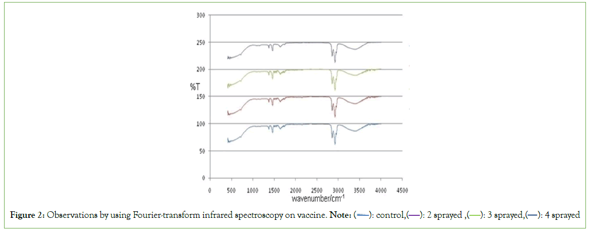
The broad peak at around 3430.86 cm-1 corresponded to the O–H stretching vibrations and the band at around 2936.93 cm-1 had the characteristic absorption of a weak C–H stretching vibration. The absorption peak at around 1620.24 cm-1 was attributed to the C=O asymmetric stretching vibration. Besides, in the region of 1000-1200 cm-1, the absorption band which was dominated by ring vibrations overlapped with the (C–O–C) glycosidic band vibrations and (C–OH) stretching vibrations of side groups. The absorption peaks at around 666 cm-1 in control are shifted to 726 cm-1 in 4 sprayed samples. The peak at 2120 cm-1 is present only in control and 2 sprayed samples which are absent in other samples. This represents the alkyne. Hence there is a lack of unsaturation in 3 and 4 sprayed samples. The absorption peaks at around 1450 cm-1 are due to methyl groups (Figure 2).
Figure 2: Observations by using Fourier-transform infrared spectroscopy on vaccine. Note: ( ): control,(
 ): 2 sprayed ,(
 ): 3 sprayed,(
 ): 4 sprayed
Proton NMR
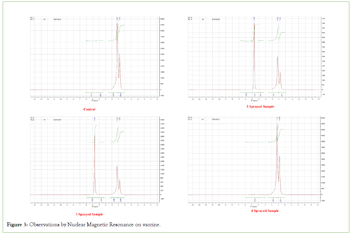
1.2 ppm-s, CH3 group 4.3-4.5 ppm-corresponding to anomeric protons of sugar moieties, 3.6-3.9 ppm-protons of sugar moieties. Also showed that the samples differ only in the point of attachment of the fatty acyl moiety to the fucose sugar ring. The isomerization involves ionization of the 3-hydroxy group and intramolecular acyl transfer from the 4-hydroxy group as the number of protons is the same in sprayed samples compared to the control. (Figure 3).
Figure 3: Observations by Nuclear Magnetic Resonance on vaccine.
Compared with control data, the instrumentation data proved that MIRGA depending on the number of sprayings had altered the chemistry of the vaccine leading to enhanced (2 sprayed)/decreased (4 sprayed) inherent characters.
Toxicological study on MIRGA: Even though MIRGA generates the safe 2-6 μm mid-IR energy, and spraying is done 0.25-0.50 meter externally right away to the packaged vaccine, we also studied the MIRGA’s toxicity effect by cytotoxicity assay [17].
Background of the MIRGA invention, MIRGA definition, techniques of mid-IR generation from MIRGA, and safety study of MIRGA using cell lines are dealt with by [17-19] (detailed discussion present in Supplementary text T2)
The action of MIRGA emitted 2-6 μm mid-IR on vaccine
While spraying MIRGA, most of the generated mid-IR energy scatters through the air and gets absorbed by vaccine molecules. Virtually all organic compounds absorb mid-IR radiation nonionizing which causes a change in the molecule’s vibrational state to move from the lower ground state to an excited higher energy state 10. This leads to changes in chemical bonds 9,10 and these bond parameter changes naturally led to consequent changes in the FMD vaccine’s physical and chemical characters, configuration and followed by compound transformation depending on the dose of energy applied [27-30].
As displayed in the results, 2-6 μm mid-IR generated from the MIRGA caused chemical and molecular level changes in the vaccine components-photodegradation (alterations of materials by light). In this process, chemical components of the vaccine have absorbed the MIR generated by MIRGA spraying, and the absorbed mid-IR photons have altered the chemical bonds of the vaccine molecule; thereby the vaccine molecules are degraded and/or transformed as reported in GCMS analysis. has explained this process as the photo dissociation of molecules caused by the absorption of photons of sunlight, such as infrared radiation, visible light, and ultraviolet light leading to a change in a molecule’s shape [31].
The laboratory analysis demonstrated that MIRGA spraying had changed the chemical bond configuration and transformation of vaccine constituents, thus increased the N-glycosylation, vaccine saturation, and isomerization and ionization and led to enhanced antibody production, especially in 2 sprayings.
All living cells’ chemical bonds’ vibrational frequencies are located in the mid-IR range [12]. The applied mid-IR is absorbed by the vaccine [32], and changes chemical bonds as shown [5,33] leading to physicochemical changes [28,30] hence enhanced antibody production.
Adjuvants are the key (stand-alone) constituent for any inactivated vaccine. At present, besides self-adjuvination and a combination of advanced techniques, chemical/biological adjuvants are used in pediatric/adult veterinary vaccine production [34,35]. Recently, four types of laser adjuvants were tested in laboratory animals successfully; the infrared laser was not used in the vaccine, but instead, applied on the patient’s skin [36]. This technique simulates the natural exposure to the sun’s radiation (sunbath) in which 66% of received solar radiation is infrared [9]. Comparatively, now MIRGA is an economical, safe method until further laser adjuvants for vaccines are evolved in full. The 2020 coronavirus pandemic was a great global challenge in developing a suitable adjuvant, which persists. However elaborate studies are ongoing at our end by using MIRGA and veterinary vaccines.
Future benefits
We found that MIRGA enhanced anti-FMDV antibodies by several folds, hence the possibility of a reduction in vaccine quantity, vaccine production cost, and host stress. Hence 30% vaccine dose may be reduced to get regular antibody titer production; or 30% extra antibody production in the sprayed regular dose.
This research could be key research for human and veterinary vaccine potentiation.
Conclusion
Mid-IR application is demonstrated to enhance the antibody quantity by several-fold in FMD vaccine. This pioneering laser adjuvant study paves the way for other vaccine/pharmaceutical potentiation. This technology can easily be applied safely from the manufacturing industry to end-users. The vaccine and adjuvant research are an endless journey. It offers savings in both money and resources, without any adverse effects. In the future, it could serve as an additional method or supplement to enhance the effectiveness of vaccines.
References
- Moni SS, Abdelwahab SI, Jabeen A, Elmobark ME, Aqaili D, Ghoal G, et al. Advancements in Vaccine Adjuvants: The Journey from Alum to Nano Formulations. Vaccines. 2023; 11(11): 1704.
- Zhao T, Cai Y, Jiang Y, Wei Y, Yu Y, Tian X, et al. Vaccine adjuvants: Mechanisms and platforms. Signal Transduct Target Ther. 2023; 8(1): 283.
- Giasuddin M, Ali M, Sayeed M, Islam E. Financial loss due to foot and mouth disease outbreak in cattle in some affected areas of Bangladesh. Bangladesh J of Livestock Res. 2021; 27(1-2): 82-94.
- Kashiwagi S, Yuan J, Forbes B, Hibert ML, Lee ELQ, Whicher L, et al. Near-Infrared Laser Adjuvant for Influenza Vaccine. PLoS ONE. 2013; 8(12):e82899.
- Tsai SR, Hamblin MR. Biological effects and medical applications of infrared radiation. J Photochem Photobiol B: Biology. 2017; 170: 197-207.
- Levy M, Bertram JR, Eller KA, Chatterjee A, Nagpal P. Nearâ?Infraredâ?Lightâ?Triggered Antimicrobial Indium Phosphide Quantum Dots. Angew Chem Int Ed Engl. 2019; 131(33):11536-11540.
- Wang F, Gao J, Wang S, Jiang J, Ye Y, Ou J, et al. Near infrared light activation of an injectable whole-cell cancer vaccine for cancer immunoprophylaxis and immunotherapy. Biomater Sci. 2021; 9(11): 3945-3953.
- CORDIS: European commission. New advances in mid-infrared laser technology, Compact, high-energy, and wavelength-diverse coherent mid-infrared source. 2015.
- Aboud SA, Altemimi AB, RS Al-HiIphy A, Yi-Chen L, Cacciola F. A Comprehensive Review on Infrared Heating Applications in Food Processing. Molecules. 2019; 24(22): 2-21.
- Girard J. Principles of Environmental Chemistry. 3rd ed. Jones & Bartlett Publishers; 2013.
- Prasad NS. Optical communications in the mid-wave IR spectral band. J Optic Comm Rep. 2005; 2(6): 558-602.
[Crossref]
- Toor F, Jackson S, Shang X, Arafin S, Yang H. Mid-infrared Lasers for Medical Applications: Introduction to the feature issue. Biomed Opt Express. 2018; 9(12): 6255.
- Grassani D, Tagkoudi E, Guo H, Herkommer C, Yang F, Kippenberg TJ, et al. Mid infrared gas spectroscopy using efficient fiber laser driven photonic chip-based supercontinuum. Nat Commun. 2019; 10(1):1553.
- Pereira MF, Shulika O, eds. Terahertz and Mid Infrared Radiation: Generation, Detection and Applications. Springer Netherlands; 2011.
- Barolet D, Christiaens F, Hamblin MR. Infrared and skin: Friend or foe. J Photochem Photobiol B: Biology. 2016; 155: 78-85.
- Nagaya T, Okuyama S, Ogata F. Near infrared photoimmunotherapy targeting bladder cancer with a canine anti-Epidermal Growth Factor Receptor (EGFR) antibody. 2018;9(27):19026-19038.
- Umakanthan, Mathi M. Decaffeination and improvement of taste, flavor and health safety of coffee and tea using mid-infrared wavelength rays. Heliyon. 2022;8(11):11338.
- Umakanthan T, Mathi M. Increasing saltiness of salts (Nacl) using midâ?infrared radiation to reduce the health hazards. Food Sci Nutr. 2023;11(6):3535-3549.
- Thangaraju U, Mathi M. Quantitative reduction of heavy metals and caffeine in cocoa using mid infrared spectrum irradiation. J Indian Chem Soc. 2023;100(1):100861.
- Dukenbayev K, Korolkov I, Tishkevich D. Fe3O4 Nanoparticles for Complex Targeted Delivery and Boron Neutron Capture Therapy. Nanomaterials. 2019;9(4):494.
- Tishkevich DI, Korolkov IV, Kozlovskiy AL. Immobilization of boron-rich compound on Fe3O4 nanoparticles: Stability and cytotoxicity. J Alloys Compd. 2019;797:573-581.
- Kozlovskiy AL, Alina A, Zdorovets MV. Study of the effect of ion irradiation on increasing the photocatalytic activity of WO3 microparticles. J Mater Sci: Mater Electron. 2021;32(3):3863-3877.
- El-Shater RE, El Shimy H. Synthesis, characterization, and magnetic properties of Mn nanoferrites. J Alloys Compd. 2022;928:166954. Google Scholar]
- Kozlovskiy AL, Zdorovets MV. Effect of doping of Ce4+/3+ on optical, strength and shielding properties of (0.5-x)TeO2-0.25MoO-0.25Bi2O3-xCeO2 glasses. Mater Chem Phys. 2021;263:124444.
- Almessiere MA, Algarou NA, Slimani Y. Investigation of exchange coupling and microwave properties of hard/soft (SrNi0.02Zr0.01Fe11.96O19)/(CoFe2O4)x nanocomposites. Materials Today Nano.
- Basagoudanavar SH, Hosamani M, Tamil Selvan RP. Development of a liquid-phase blocking ELISA based on foot-and-mouth disease virus empty capsid antigen for seromonitoring vaccinated animals. Arch Virol. 2013;158(5):993-1001.
- Atkins P, Paula J de. Physical Chemistry for the Life Sciences. OUP Oxford. 2011.
- Yi GC. Semiconductor Nanostructures for Optoelectronic Devices: Processing, Characterization and Applications. Springer Berlin Heidelberg. 2012.
- Datta SN, Trindle CO, Illas F. Theoretical and Computational Aspects of Magnetic Organic Molecules. Imperial college press. 2014.
- Esmaeili K. Viremedy. Homeopathic remedies, energy healing remedies as information including remedies, a synopsis. 2023. [Crossref]
- Yousif E, Haddad R. Photodegradation and photostabilization of polymers, especially polystyrene: review. SpringerPlus. 2013;2(1):398.
- Sommer AP, Zhu D, Mester AR, Forsterling HD. Pulsed Laser Light Forces Cancer Cells to Absorb Anticancer Drugs-The Role of Water in Nanomedicine. Artif Cell Blood Sub. 2011;39(3):169-173.
- Mohan J. Organic Spectroscopy: Principles and Applications, 2nd Edition. Alpha Science International Ltd, Harrow, UK. 2004.
- Bonam SR, Partidos CD, Halmuthur SKM, Muller S. An Overview of Novel Adjuvants Designed for Improving Vaccine Efficacy. Trends Pharmacol Sci. 2017;38(9):771-793.
- Nanishi E, Dowling DJ, Levy O. Toward precision adjuvants: Optimizing science and safety. Curr Opin Pediatr. 2020;32(1):125-138.
- Kashiwagi S. Laser adjuvant for vaccination. FASEB j. 2020;34(3):3485-3500.
Citation: Umakanthan T, Mathi M, Devi U, Ramakrishnan S (2024) Potentiation of Foot and Mouth Disease Vaccine by Irradiating Mid-Infrared Rays-A Laser Adjuvant Technology. J Vaccines Vaccin. 15:558.
Copyright: © 2024 Umakanthan T, et al. This is an open-access article distributed under the terms of the Creative Commons Attribution License, which permits unrestricted use, distribution, and reproduction in any medium, provided the original author and source are credited.


