
Indexed In
- RefSeek
- Hamdard University
- EBSCO A-Z
- Publons
- Euro Pub
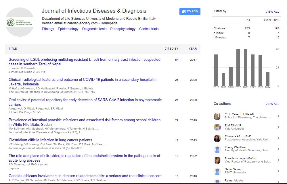
- Google Scholar
Useful Links
Share This Page
Journal Flyer
Open Access Journals
- Agri and Aquaculture
- Biochemistry
- Bioinformatics & Systems Biology
- Business & Management
- Chemistry
- Clinical Sciences
- Engineering
- Food & Nutrition
- General Science
- Genetics & Molecular Biology
- Immunology & Microbiology
- Medical Sciences
- Neuroscience & Psychology
- Nursing & Health Care
- Pharmaceutical Sciences
Commentary - (2024) Volume 9, Issue 6
Plague: Diagnosis, Clinical Manifestations and Modern Approaches to Detection
Rafael Jose*Received: 28-Oct-2024, Manuscript No. JIDD-24-27709; Editor assigned: 30-Oct-2024, Pre QC No. JIDD-24-27709 (PQ); Reviewed: 14-Nov-2024, QC No. JIDD-24-27709; Revised: 21-Nov-2024, Manuscript No. JIDD-24-27709 (R); Published: 29-Nov-2024, DOI: 10.35248/2576-389X.24.09.301
Description
Plague, a zoonotic disease caused by Yersinia pestis, remains a significant public health concern despite advancements in medicine. Historically responsible for devastating pandemics such as the Black Death, it persists today in endemic regions. Transmission occurs through fleas, direct contact, or aerosols, with three primary clinical forms: Bubonic, septicemic and pneumonic. Rapid diagnosis and treatment are essential to reduce mortality. Plague, caused by Yersinia pestis, is a highly virulent bacterial infection that has shaped human history. Found predominantly in rodent populations, it is transmitted to humans through flea bites, contact with infected animals, or inhalation of respiratory droplets. Though less prevalent today, outbreaks occur in rural and semi-rural areas of Africa, Asia and the Americas, where animal reservoirs persist. With its rapid disease progression and potential for severe outcomes, accurate diagnosis remains a priority for public health authorities.
Etiology and pathophysiology
Yersinia pestis is a gram-negative coccobacillus belonging to the Enterobacteriaceae family. It possesses several virulence factors that allow it to evade immune responses, including type III secretion systems and antiphagocytic proteins. The bacteria can proliferate in lymph nodes, causing inflammation and necrosis, characteristic of bubonic plague. In advanced stages, it may enter the bloodstream, leading to septicemia, or disseminate to the lungs, causing pneumonic plague.
Clinical manifestations
Plague presents in three primary clinical forms, each with distinct features:
Bubonic plague: The most common form, characterized by sudden fever, chills and painful swollen lymph nodes (buboes). Without treatment, it can progress to septicemia.
Septicemic plague: Results from untreated bubonic plague or direct entry of bacteria into the bloodstream. Symptoms include fever, abdominal pain and disseminated intravascular coagulation, which can lead to necrosis and gangrene.
Pneumonic plague: The most severe form, arising from primary inhalation of infected aerosols or secondary spread from septicemia. Symptoms include acute respiratory distress, hemoptysis and high mortality without prompt antibiotic therapy.
Diagnostic methods
Timely and accurate diagnosis is vital to manage plague effectively. Various diagnostic tools are available, each with specific applications:
Clinical assessment: Diagnosis often begins with identifying characteristic symptoms such as sudden fever and buboes. However, clinical signs alone are insufficient due to overlap with other febrile illnesses.
Microscopy: Direct examination of specimens (e.g., lymph node aspirates, blood, or sputum) using Gram stain or Wright-Giemsa stain can reveal bipolar-staining Y. pestis. Though rapid, this method lacks specificity.
Polymerase Chain Reaction (PCR): PCR-based methods detect Y. pestis DNA in clinical specimens with high sensitivity and specificity. These techniques allow for rapid confirmation, often within hours.
Serological tests: Antibody detection in paired serum samples using Enzyme-Linked Immunosorbent Assay (ELISA) or passive hem-agglutination tests provides retrospective confirmation. Serological methods are less useful for acute diagnosis due to the time required for antibody production.
Rapid Diagnostic Tests (RDTs): Immunochromatographic assays targeting the F1 antigen of Y. pestis offer point-of-care testing options in resource-limited settings. These tests are easy to use and provide results within minutes but may lack accuracy in some cases.
Advanced imaging: Radiographic imaging can aid in identifying complications, such as pulmonary involvement in pneumonic plague. While not diagnostic, imaging supports clinical evaluation.
Challenges in diagnosis
Diagnosing plague is complicated by its overlapping symptoms with other febrile illnesses, such as typhoid fever or leptospirosis. Additionally, limited laboratory capacity in endemic regions may delay confirmation. Strengthening local diagnostic capabilities and enhancing disease surveillance are essential to address these challenges.
Treatment
Prompt antibiotic therapy significantly improves survival rates. Streptomycin and gentamicin are the drugs of choice, with doxycycline and ciprofloxacin as alternatives. Supportive care, including fluid management and oxygen therapy for pneumonic cases, is also important.
Prevention and control
Preventing plague involves reducing human exposure to infected rodents and fleas. Public health measures include rodent control, use of insect repellents and educating at-risk populations. Vaccines under development show potential for long-term prevention in high-risk areas.
Conclusion
Plague continues to pose a threat in endemic regions and requires vigilance due to its rapid progression and potential for outbreaks. A combination of clinical suspicion, laboratory diagnostics and timely treatment is essential to mitigate its impact. Strengthening diagnostic capacity and public health systems in affected areas will improve outcomes and prevent future epidemics.
Citation: Jose R (2024). Plague: Diagnosis, Clinical Manifestations and Modern Approaches to Detection. J Infect Dis Diagn. 9:301.
Copyright: © 2024 Jose R. This is an open-access article distributed under the terms of the Creative Commons Attribution License, which permits unrestricted use, distribution and reproduction in any medium, provided the original author and source are credited.