Indexed In
- Open J Gate
- Genamics JournalSeek
- JournalTOCs
- Ulrich's Periodicals Directory
- RefSeek
- Hamdard University
- EBSCO A-Z
- OCLC- WorldCat
- Proquest Summons
- Publons
- Geneva Foundation for Medical Education and Research
- Euro Pub
- Google Scholar
Useful Links
Share This Page
Journal Flyer

Open Access Journals
- Agri and Aquaculture
- Biochemistry
- Bioinformatics & Systems Biology
- Business & Management
- Chemistry
- Clinical Sciences
- Engineering
- Food & Nutrition
- General Science
- Genetics & Molecular Biology
- Immunology & Microbiology
- Medical Sciences
- Neuroscience & Psychology
- Nursing & Health Care
- Pharmaceutical Sciences
Case Report - (2021) Volume 12, Issue 5
Oesophageal Carcinosarcoma a Rare Neoplasm of the Oesophagus
Amaresh Aruni*Received: 09-Jun-2021 Published: 30-Jun-2021, DOI: 10.35248/2155-9864.21.12.466
Abstract
Oesophageal carcinosarcoma is a rare oesophageal cancer, expressing both carcinomatous and sarcomatous elements. We report a 60-year male presenting with progressive dysphagia and endoscopic biopsy revealed leiomyosarcoma and underwent minimally invasive esophagectomy, gastric conduit and cervical anastomosis, final histopathology revealed carcinosarcoma. Post-operatively received adjuvant chemo radiation and at 3 months follow-up developed dysphagia due to anastomotic stricture managed with endoscopic dilatation. At 12 months follow up patient is asymptomatic and no radiological evidence of recurrence.
Keywords
Carcinosarcoma; Leiomyosarcoma; Adenocarcinoma
Introduction
Oesophageal cancer is the third most common gastrointestinal cancer. Oesophageal adenocarcinoma and squamous cell carcinoma are the most common oesophageal cancers. Oesophageal carcinosarcoma is rare malignant neoplasm of the oesophagus, expressing both carcinomatous and sarcomatous elements. Due to rarity of the lesion many controversies exist regarding the management. We report a rare case of oesophageal carcinosarcoma managed with surgery (minimally invasive esophagectomy, gastric conduit and cervical anastomosis) and adjuvant chemo radiation [1].
Case Representation
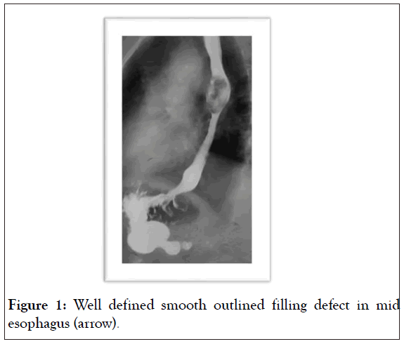
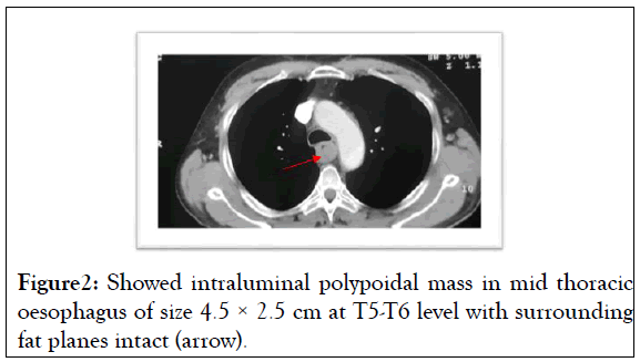
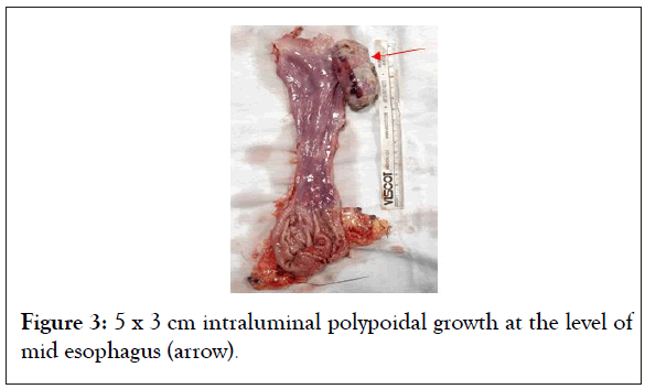
60-year male with history of chronic smoking and alcohol use, presented with progressive dysphagia for 4 months, no history of hematemesis, odynophagia and no family history of similar complaints. Clinical examination findings were normal, no lymphadenopathy. Evaluated with UGIE revealed- polypoidal intraluminal growth at mid oesophagus with significant luminal narrowing and endoscopic biopsy was suggestive of Leiomyosarcoma [2]. On barium swallow- well defined smooth outlined filling defect in mid oesophagus-5.4 × 3.5 cm). CECT chest showed intraluminal polypoidal mass in mid thoracic oesophagus of size 4.5 × 2.5 cm at T5-T6 level with surrounding fat planes intact. Underwent minimally invasive esophagectomy with gastric conduit and cervical oesophago-gastric anastomosis. 5 × 3 cm intraluminal polypoidal growth at the mid oesophagus [3]. Post-operative period had gastroparesis for 7 days, managed with prokinetics and discharged on post-op day 15. Histopathology turned out to be carcino-sarcoma. Received 45 by of radiotherapy and 6 cycles of cisplatin and 5FU, no side effects noted [4]. After follow up of 3 months developed grade 2 dysphagia due to anastomotic stricture, improved with endoscopic dilatation in two sittings. At 10 months follow up, patient is asymptomatic, with no clinical and radiological evidence of recurrence (Figure 1-3).

Figure 1: Well defined smooth outlined filling defect in mid esophagus (arrow).
Figure 2: Showed intraluminal polypoidal mass in mid thoracic oesophagus of size 4.5 × 2.5 cm at T5-T6 level with surrounding fat planes intact (arrow).
Figure 3: 5 x 3 cm intraluminal polypoidal growth at the level of mid esophagus (arrow).
Discussion
Oesophageal carcinoma are epithelial tumours constitute majority of the oesophageal neoplasms. Esophageal cancer ranks 7th in incidence and 6th most common cause of cancer related death. Oesophageal carcinosarcoma is rare oesophageal neoplasm (0.5-1.5%) expressing both squamous and sarcomatous elements, first named by virchow in 1865 and first reported. Esophageal carcinosarcoma is also called as pseudo sarcoma, spindle cell carcinoma, sarcomatoid carcinoma. Esophageal carcinosarcoma is more prevalent in men, especially with history of tobacco and alcohol use.
The reason for oesophageal carcinosarcoma is hypothesized in to three theories: metaplastic theory (spindle cell carcinoma)-there is a single stem cell, and through metaplasia of original squamous cell carcinoma, the sarcomatous component arises. Collision theory (true carcinosarcoma)- there are two types of stem cell each differentiates in to carcinomatous and sarcomatous cells. Epithelial tumour theory (pseudo sarcoma), with a mesenchymal component that is non-neoplastic in origin, instead representing a reactive process to the presence of the epithelial neoplasm.
The presentation of oesophageal carcinosarcoma is almost similar but symptoms are rapidly progressive due to faster growth of the tumour, and symptoms are dysphagia (80%), odynophagia, weight loss, anorexia.
The oesophageal carcinosarcoma most commonly located in the middle third of the oesophagus (59%), followed by lower one third (28%), upper one third (13%). Endoscopic findings are polypoidal growth with smooth surface and causing luminal narrowing difficult to differentiate the type of tumour. Macroscopically, carcinosarcomas are two types polypoid type (75%) seen when sarcomatoid component is predominant and ulcerative type (15%) when carcinomatous component is predominant [5].
On barium studies reveals cupola sign, which refers to the dome like intraluminal filling defect with smooth outline surface helps to predict the possibility of the mural origin of the oesophageal tumour. It is difficult to distinguish the pathology of tumour on radiological investigations. Other investigations applicable are CECT and EUS, EUS helps to diagnose the T stage of the tumour with high sensitivity. Furthermore, endoscopic biopsy only captures the superficial aspect of the mass, often showing the squamous cell component but not the deeper sarcomatous cell component. Hence for the final diagnosis requires the entire resected specimen. In our case the endoscopic biopsy diagnosis was only leiomyosarcoma but surgical resected specimen turned out to be carcinosarcoma, probably the primary lesion was ulcerated which failed to take epithelial component in endoscopic biopsy. The characteristics of carcinosarcoma in pathology are both squamous cell component, stain positively for cytokeratin. The sarcomatous component shows spindle cells as fascicles or whorled pattern and stain positive.
Due to rare incidence, management of oesophageal carcinosarcoma has many controversies. Majority of tumours present as polypoidal type with less deep invasive than oesophageal carcinoma. Reported the percentage of T1/2 lesions was higher in the oesophageal carcinosarcoma group than in the oesophageal squamous cell carcinoma group. Hence endoscopic management can be considered for early stage tumours. However the management of oesophageal carcinosarcoma with surgical resection (partial or total esophagectomy) with regional lymph nodal dissection is better because the pre-operative rate of diagnosis of oesophageal carcinosarcoma is very low, risk of regional lymph node metastasis is around 50-67% as comparable to oesophageal squamous cell carcinoma, high-grade hyperplasia or carcinoma in situ was found in adjacent oesophageal mucosa at a certain distance from the tumour body and lastly the possibility of remnant tumour in area of previous ECS after endoscopic treatment [6].
Esophageal carcinosarcoma appear to have better prognosis than oesophageal carcinoma, because these tumours have high tendency to grow intraluminally, symptomatic at early stage and incidence of lymph nodal metastasis is thus low. However, 5-year survival rates are comparable following treatment. Compared 20 cases of carcinosarcoma with 773 cases of squamous cell carcinoma. Although the 3-year survival rate was higher for carcinosarcoma (62.8%) than for squamous cell carcinoma (28.1%), no significant difference in 5-year survival is apparent between the two groups.
Conclusion
Neoadjuvant chemotherapy regimen 5FU and cisplatin helps in achieving the complete response or partial response, increases the disease-free survival after surgery. Chemo radiotherapy may be effective definitive therapy for cervical ECS. In our case neoadjuvant was not used as the endoscopic biopsy was suggestive of leiomyosarcoma. Capecitabine, docetaxel, platinum based chemotherapeutic agents and radiotherapy are used to decrease the local and distant recurrence. Molecular targeted therapy against MET and EGFR may be effective in inhibiting the growth or progression of the epithelial and/or mesenchymal component of ECS. Further studies are required to know the exact efficacy of the molecular targeted therapy.
Summary
Although esophageal carcinosarcoma is rare neoplasm of the esophagus. Neoadjuvant chemotherapy helps in achieving pathological response. Esophageal carcinosarcoma have better prognosis than esophageal carcinoma. Surgery is the treatment of choice.
REFERENCES
- Xu X, Xu Y, Wang J, Zhao C, Liu C, Wu B, et al. The controversy of esophageal carcinosarcoma: A case report and brief review of literature. Medicine (Baltimore). 2019;98(10):e14787.
- Japan Esophageal Society. Japanese classification of esophageal cancer, 11th Edition: part II and III. Esophagus. 2017;14(1):37–65.
- Bray F, Ferlay J, Soerjomataram I, Siegel RL, Torre LA, Jemal A, et al. Global cancer statistics 2018: GLOBOCAN estimates of incidence and mortality worldwide for 36 cancers in 185 countries. CA Cancer J Clin. 2018;68(6):394–424
- Au JT, Sugiyama G, Wang H, Nicastri A, Lee D, Ko W, et al. Carcinosarcoma of the oesophagus – a rare mixed type of tumor. J Surg Case Rep. 2010;2010(7):7–7.
- Agha F, Keren D. Spindle-cell squamous carcinoma of the esophagus: a tumor with biphasic morphology. Am J Roentgenol. 1985;145(3):541–5.
- Sano A, Sakurai S, Kato H, Suzuki S, Yokobori T, Sakai M, et al. Expression of receptor tyrosine kinases in esophageal carcinosarcoma. Oncol Rep. 2013;29(6):2119–26.
Citation: Aruni A, (2021) Oesophageal Carcinosarcoma A Rare Neoplasm of the Oesophagus. J Blood Disord Transfus. 12:1000466.
Copyright: © 2021 Aruni A. This is an open-access article distributed under the terms of the Creative Commons Attribution License, which permits unrestricted use, distribution, and reproduction in any medium, provided the original author and source are credited.


