Indexed In
- Open J Gate
- RefSeek
- Hamdard University
- EBSCO A-Z
- OCLC- WorldCat
- Publons
- Geneva Foundation for Medical Education and Research
- Euro Pub
- Google Scholar
Useful Links
Share This Page
Journal Flyer
Open Access Journals
- Agri and Aquaculture
- Biochemistry
- Bioinformatics & Systems Biology
- Business & Management
- Chemistry
- Clinical Sciences
- Engineering
- Food & Nutrition
- General Science
- Genetics & Molecular Biology
- Immunology & Microbiology
- Medical Sciences
- Neuroscience & Psychology
- Nursing & Health Care
- Pharmaceutical Sciences
Case Report - (2023) Volume 12, Issue 6
Multiple Pulmonary Hydatid Cysts with Bronchial Communication Imitating as Tuberculosis
Kamal Garg*, Sunita Aggarwal, Abhishek Verma, Tushar Mittal, Ranvijay Singh and Dhananjay M KharcheReceived: 23-Oct-2023, Manuscript No. CMO-23-23655; Editor assigned: 25-Oct-2023, Pre QC No. CMO-23-23655(PQ); Reviewed: 08-Nov-2023, QC No. CMO-23-23655; Revised: 15-Nov-2023, Manuscript No. CMO-23-23655(R); Published: 23-Nov-2023, DOI: 10.35248/2327-5073.23.12.371
Abstract
Hydatid disease is a common disease which is usually asymptomatic, can present as dull pain or pressure symptoms. Very rarely, it presents as a febrile illness with pulmonary cysts. We present here a case of an 18 year old girl who presented with fever and productive cough since 1 month with an episode of hemoptysis. In our country, tuberculosis is kept as the first differential. Investigations revealed a cystic lesion with detached membrane within, with normal lung parenchyma and no lymphadenopathy. The patient was initially started on anti-parasitic therapy. Later on, the patient underwent video assisted thoracic surgery following which the patient improved clinically within a few days.
Keywords
Pulmonary cysts; Anti-parasitic therapy; Tuberculosis
Introduction
Hydatid disease is zoonotic parasitic infection caused by larval stage of Echinococcus sp. Life cycle consists of dogs being the definitive host who harbour the adult tapeworms and herbivores as intermediate host who develop hexacanth embryo and cysts. Humans are accidental host through contaminants like soiled foods [1-4].
The disease is generally asymptomatic, usually presents clinically as dull pain or pressure on corresponding organs. The liver is involved in about two-thirds of E. granulosus infections and in nearly all E. multilocularis infection. Lung involvement is seen in ~18% of cases, usually as lung cyst which may or may not be symptomatic [5,6]. Diagnosis is made with high clinical suspicion, serology and radiological investigations.
The common causes of symptomatic pulmonary cysts in our country are benign cyst, infective cyst, tubercular abscess and malignant cyst [3]. Our case highlights the need for screening of hydatid infection in such patients. As pulmonary cyst patients are uncommon, it is crucial to differentiate between presentations of isolated pulmonary multiple cysts with hydatid as the aetiology. The majority of cases are discovered accidentally, therefore disease awareness among the general public and among healthcare professionals is essential forprevention, early management, and the reduction of consequences.
Case Presentation
We report here a case of an adolescent female who presented with complaints of fever and pain on right side of the chest since 1 month and cough since 2 weeks. The fever was documented, 101 F, insidious in onset, remittent in nature, associated with evening rise, not associated with chills or rigors and relieved by anti pyretics. Her pain was dull aching in nature associated with heaviness on same side. Cough was associated with sputum, which was mucoid in nature and also had an episode of hemoptysis. She had no complaints of dyspnea, anorexia and weight loss. There was no history of trauma to the chest or palpitations. She had no significant past history of occupational exposure, allergy, any surgery, active tuberculosis contact and any drug use. Her family owns a dairy farm along with a pet dog. On examination, patient had a BMI of 22.6 kg/m2 with no pallor or icterus. There was no lymphadenopathy. She was vitally stable with body temperature 99.9ºF in axilla. Her chest examination revealed reduced air entry with coarse crepitations in right infra- axillary and infra-scapular areas, with rest systemic examination being normal.
Investigations
Laboratory analysis demonstrated a normocytic anaemia with haemoglobin levels of 10.7 g/dl with total leukocyte counts of 6800/mm3 and platelet of 390,000/mm3. ESR was 64 mm/hr along-with normal liver and renal function test. Her serum procalcitonin was 0.17 ng/ml, hsCRP was 3 mg/L.
Her ECG revealed normal sinus rhythm. Her blood and urine cultures were sterile. Her serology for HIV, HBV and HCV were non reactive. Tuberculin skin test was 3 mm at the end of 48 hrs.
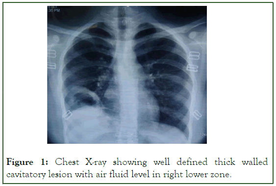
Acid fast bacilli, fungal hyphae or any bacteria were not detected on serial sputum investigations. Her ANA/ ANCA profile, Rheumatoid factor and CRP were non reactive. A chest skiagram reveals a right lower zone thick cavitatory lesion with air fluid level within (Figure 1).

Figure 1: Chest X-ray showing well defined thick walled cavitatory lesion with air fluid level in right lower zone.
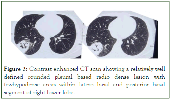
An ultrasonography of local site reveals cystic lesion with detached membranes in right lower lung lobe along-with air with probable bronchial communication. Radiographs and sonography of appendicular skeleton and musculature revealed no other cystic or calcified lesion. Contrast enhanced chest and abdominal computed tomography revealed well defined round hypo-dense collection of 4.5 × 4.8 × 4.2 cm in lateral and posterior basal segment of right lower lung lobe with broad based towards pleura with thick enhancing wall and few enhancing septa within (Figure 2).

Figure 2: Contrast enhanced CT scan showing a relatively well defined rounded pleural based radio dense lesion with fewhypodense areas within latero basal and posterior basal segment of right lower lobe.
No other cystic lesion found in any other organ including liver. Serology for Echinococcus was sent which came out to be positive for IgM (1:128) and IgG (1:64).
With this clinical presentation and investigations, the patient was diagnosed as a case of pulmonary hydatid cyst with probable bronchial communication.
Treatment
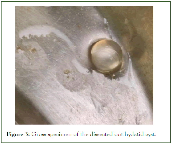
The patient was started on Tab Albendazole 400 mg twice daily basis of clinical and USG findings. Further, video assisted thoracic surgery guided evacuation of right hydatid cyst with repair of broncho-pleural communication with ICD insertion was done. Intraoperative findings reveals ruptured lung hydatid cyst with multiple other small cysts along-with broncho-pleural fistula and adhesion of cyst wall with parietal pleura (Figure 3).

Figure 3: Gross specimen of the dissected out hydatid cyst.
During dissection, multiple membranes along with multiple fully intact cyst was extracted. Postoperative, ICD output monitoring was done which was eventually removed after 3 days. There was no output in the ICD bag. Patient was put on month long Albendazole therapy.
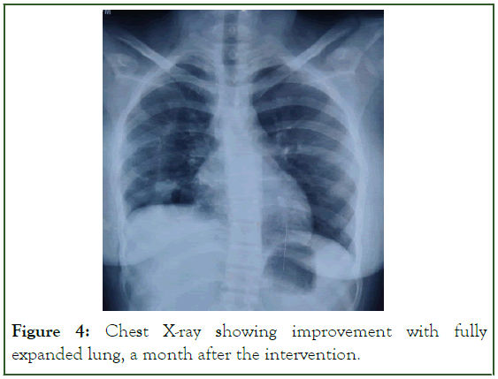
The patient improved symptomatically and became ambulatory after ICD removal. Within a week, patient’s fever and cough improved. After 4 weeks duration, repeat chest radiograph was taken which revealed no lesions left and fully expanded lung parenchyma (Figure 4).

Figure 4: Chest X-ray showing improvement with fully expanded lung, a month after the intervention.
Drug therapy continued for a duration of 1 month [7]. Repeat ultrasonography revealed no cystic lesions left. Patient returned to her routine self. She is on routine follow up and doing completely fine.
Results and Discussion
Hydatid disease is caused by Echinococcus sp., especially E. granulosus and E. multilocularis. The disease spreads with ingestion of food contaminated with faeces of the definitive host. The disease is mostly seen in villages where sheep and dogs are in close contacts with the humans [3,4]. The liver followed by lung are the most common site [5,6].
The mean age of presentation is around 4th decade [8]. The clinical manifestations of the disease vary with the size and compressive symptoms on the particular organs [5]. Isolated presence of multiple pulmonary cyst with bronchial communication mimicking as tuberculosis is extremely rare. Intraoperative presence of multiple cyst in a radiologically single defined cyst is also very rare.
Due to indolent course of the disease and long asymptomatic period, the pathology is not easily picked up. Also, there are no standard diagnostic guidelines and resources are lacking in disease prevalent rural areas. Long standing fever with productive cough in our case highlights the role of hydatid disease as a possible aetiology in patients presenting with pulmonary cyst.
Chest radiographs, CT and serology are commonly used diagnostic modalities [8]. Inclusion of sonography in diagnosis seems to be of paramount importance because of the ability to visualize membranes. Excision followed by microscopic diagnosis is considered as standard.
The medical treatment generally comprises of anti-helminthic drugs. Surgical management is the main stay of treatment, with either placement of ICD tube under steroid cover followed by surgical removal of remaining membranes or surgical removal with maximum possible sparing of lung parenchyma [9,10].
Our case shows possibility of Hydatid disease as an important differential in a patient with pulmonary cyst. In our case, the patient underwent a surgical removal with repair. It shares clinical picture with many differentials. Pneumonia, benign pulmonary cyst, infected pulmonary cyst and neoplasm are the important differentials. Low degree of suspicion makes it a diagnostic challenge. Focusing on deworming of dogs can be possible to contain the disease. Slaughterhouse hygiene, public education and consumption of properly cooked meals should also be promoted.
Conclusion
Hydatid cyst is a relatively indolent disease, but should be suspected in case of lung cyst. The disease has variable presentation and shares clinical picture with many other diseases like tuberculosis. Anti-parasitics are used, though surgery being the main stay of the treatment carries a high risk of rupture and anaphylaxis. The disease can be easily controlled with consumption of appropriately washed and well cooked food with proper hygiene.
References
- Ramos G, Md P, Antonio Orduña M, Mariano García-yuste M. Hydatid cyst of the lung: Diagnosis and treatment. World J Surg. 2001;25(1):46.
[Crossref] [Google Scholar] [PubMed]
- Marla NJ, Marla J, Kamath M, Tantri G, Jayaprakash C. Primary hydatid cyst of the lung: A review of the literature. J Clin Diagnostic Res. 2012;6(7).
- Shukla S, Singh SK, Pujani M. Multiple disseminated abdominal hydatidosis presenting with gross hydatiduria: A rare case report. Indian J Pathol Microbiol. 2009;52(2):213.
[Crossref] [Google Scholar] [PubMed]
- Basavana GH, Siddesh G, Jayaraj BS, Krishnan MG. Ruptured hydatid cyst of lung. J Assoc Physicians India. 2007;55:141-145.
[Google Scholar] [PubMed]
- Zaman K, Mewara A, Kumar S, Goyal K, Khurana S, Tripathi P, et al. Seroprevalence of human cystic Echinococcosis from North India (2004–2015). Trop Parasitol. 2017;7(2):103.
[Crossref] [Google Scholar] [PubMed]
- Safioleas M, Stamoulis I, Theocharis S, Moulakakis K, Makris S, Kostakis A. Primary hydatid disease of the gallbladder: A rare clinical entity. J Hepatobiliary Pancreat Sug. 2004;11:352-356.
- Moro P, Schantz PM. Echinococcosis: A review. Int J Infect Dis. 2009;13(2):125-133.
[Crossref] [Google Scholar] [PubMed]
- Ali R, Sajad R, Sina S. Surgical treatment of pulmonary hydatid cystin in children. Tanaffos. 2009;8(1):56-61.
- Balci AE, Eren N, Evval Eren S, Ulku R. Ruptured hydatid cysts of the lung in children: Clinical review and results of surgery. Ann Thorac Surg. 2002;74(3):889-892.
[Crossref] [Google Scholar] [PubMed]
- Lodhia J, Herman A, Philemon R, Sadiq A, Mchaile D, Chilonga K. Isolated Pulmonary hydatid cyst: A rare presentation in a young Maasai boy from northern Tanzania. Case Rep Surg. 2019;2019.
[Crossref] [Google Scholar] [PubMed]
Citation: Garg K, Aggarwal S, Verma A, Mittal T, Singh R, Kharche DM (2023) Multiple Pulmonary Hydatid Cysts with Bronchial Communication Imitating as Tuberculosis. Clin Microbiol. 12:371.
Copyright: © 2023 Garg K, et al. This is an open access article distributed under the terms of the Creative Commons Attribution License, which permits unrestricted use, distribution, and reproduction in any medium, provided the original author and source are credited.
