Indexed In
- Academic Journals Database
- Open J Gate
- Genamics JournalSeek
- JournalTOCs
- China National Knowledge Infrastructure (CNKI)
- Scimago
- Ulrich's Periodicals Directory
- RefSeek
- Hamdard University
- EBSCO A-Z
- OCLC- WorldCat
- Publons
- MIAR
- University Grants Commission
- Geneva Foundation for Medical Education and Research
- Euro Pub
- Google Scholar
Useful Links
Share This Page
Open Access Journals
- Agri and Aquaculture
- Biochemistry
- Bioinformatics & Systems Biology
- Business & Management
- Chemistry
- Clinical Sciences
- Engineering
- Food & Nutrition
- General Science
- Genetics & Molecular Biology
- Immunology & Microbiology
- Medical Sciences
- Neuroscience & Psychology
- Nursing & Health Care
- Pharmaceutical Sciences
Review Article - (2023) Volume 14, Issue 2
Impact of Immunologic Discoveries on the Vaccine
Zelalem Kiros BitsueReceived: 22-Dec-2022, Manuscript No. JVV-22-19430; Editor assigned: 26-Dec-2022, Pre QC No. JVV-22-19430 (PQ); Reviewed: 11-Jan-2023, QC No. JVV-22-19430; Revised: 18-Jan-2023, Manuscript No. JVV-22-19430 (R); Published: 27-Jan-2023, DOI: 10.35248/2157-7560.23.14.515
Abstract
The spike glycoprotein (S1) interacts with host cell epithelial Angiotensin-Converting Enzyme-2 (ACE-2) receptors. The nano-luciferase-based assay shows that the virus’s 1 protein has a strong binding affinity with the ACE-2 receptors. Reports illustrate the S-protein antigenic epitope of SARS-CoV-2 binds with the TLR4/ MD-2 complex by strong molecular bonding interactions. The pool of miRNA based studies shows that TMPRSS2 acts as promising regulators for the SARS-CoV-2 entry checkpoint. Recent report stated that the AT1R phosphorylates JAK2 in the lung cells, which activates STAT-3 transduction to the nucleus.
Not only I discovered vaccine AZEE CRA 01, immune cell principles and methods of vaccination but also B cells have two components. B cells synthesis both MHC I, and MHC II. CD4 interacts with MQ, MHC II, CD8 Interacts with DC, MHC I, and B cells interacts with both MHC I, and MHC II, and Th2 and Tc1 (CD4 and CD8).
In these original articles I report the pandemics and immune cells, immunologic clearance and immune mechanisms, the discovery process: innovation and immunology, zelalem kiros bitsue and his vaccine discoveries, zelalem kiros bitsue and his immunologic discoveries, vaccine discovered and mechanisms of action 1of 2.
Keywords
Immune mechanisms; Immune cells; Vaccine discoveries; Pandemic; Vaccination
Introduction
Viruses have countless mechanisms to escape from the host’s immune system. To illustrate the most common strategies of viral immune escape: speed shape change, biochemical properties and pH. Multiple viruses with RNA-based genomes, often much smaller, which also manage to set up persistent infection, and survive within hosts in the face of ongoing immune responses.
Unlike their more stable DNA counterparts, the mutability of these RNA genomes allows this group, potentially, to evolve within their host, and to set up high level persistence. There is an overlap here between mechanisms of viral persistence and immune escape.
There are three phases of immune responses against replicating pathogens such as viruses. Firstly, immune induction, followed by an effector phase, and finally a prolonged state of memory.
Chimera Virus (SARS-COV-2)
Chimera virus (SARS-COV-2) mutation continue BA 2.75, BA 2.75.2, BA 4.6, BF7, BQ 1, BQ 1.1 and XBB, no vaccine yet stop chimera virus (SARS-COV-2) from mutation, replications, and spread.
One virus contains the genetic material derived from two or more distinct virus, known as chimera virus. A new hybrid microorganism created by joining nucleic acid (RNA and DNA), fragments from two or more different microorganisms.
In which each of at least two of the fragments, contains essential genes necessary for replication. This virus can potentially replicate 40%-80%.This virus can potentially mutate, replicate, and contagious.
Chimera virus (SARS-COV-2) has given us very good lesson, showed how global health system have been disorganized and mis- shapes, how global communities suffered and also global leaders frustrated.
The Pandemics and Immune Cells
SARS-CoV-2 viral entry is initiated by binding RBD of the S protein to the human host cell receptors at the cell surface [1-3]. A recent report demonstrated that spike protein had been O-glycosylated on the amino acid threonine (T678) adjacent to the furin cleavage site. Liquid chromatography-mass spectrometry analysis showed that the spike protein’s LacdiNAc structural motifs and polyLacNAc structures [4-6]. The spike glycoprotein (S1) interacts with host cell epithelial Angiotensin-Converting Enzyme 2 receptors (ACE2) [7,8]. At the N terminus of this bridge-shaped structure, Q498, T500, and N501 residues of the RBD protein form a hydrogen- bond network with Y41, Q42, K353, and R357 amino acid residues of the ACE2 peptidase domain [9]. At the C terminus of bridge- shaped α1 helix RBD Q474 forms a hydrogen bond with the ACE2 Q24, with RBD F486 interacting with the ACE2 M82 through Van der Waals forces. ACE2 receptor which is highly expressed in human epithelial, endothelial, renal, cardiovascular tissue and lung parenchyma [10]. A report suggests that SARS-CoV-2 also capable to interact with SARS-CoV-2 host targets like ACE2, CD26, Ezrin and Cyclophilins [11]. Reports illustrate the S-protein antigenic epitope of SARS-CoV-2 binds with the TLR4/ MD-2 complex by strong molecular bonding interactions [12]. The human Trans- Membrane Protease Serine 2 (TMPRSS2) processes the viral spike protein and exposes fusion peptide present in the S2 subunit to the host receptor ACE-2 [13]. This S protein processing and priming by the TMPRSS2 is an essential step in the SARS-CoV-2 infection [14]. Then, the SARS-CoV-2 uses cysteine proteases like Cathepsin B and L (CatB/L) and promotes virus-plasma membrane fusion [15,16].
Moreover, SARS-CoV-2 down-regulates the expression of ACE-2 resulted in the up-regulated expression pattern of Ang II. Ang II is formed by the degradation of Ang I by the enzyme ACE-2 [17]. The overexpressed Ang II binds with its plasma membrane receptor AT1R. This membrane-bound ATIR transduces the signals to the inflammatory transcription factors like NF-ƙB which mediates several inflammatory cytokines’ activation and overexpression [18, 19]. Further, recent report stated that the AT1R phosphorylates JAK2 in the lung cells, which activates STAT-3 transduction to the nucleus [20]. The STAT-3 is a signal transducer and activator of transcription, which initiates the active transcription of inflammatory cytokines. Additionally, the Ang II/AT1R interaction activates macrophages to produce excessive inflammatory cytokines and further contribute to “cytokine storm” and the development of Acute Respiratory Distress Syndrome (ARDS) [21-23].
Immunologic Clearance and Immune Mechanisms
T cell independent B cell activation
The antigen which triggers B cell activation without T cell help; clustering of BCR activates multiple Igα/Igβ molecules and initiates the signaling process clustering B cell requires activating accessories proteins Igα/Igβ Signal 1: Clustering B cell receptor depends of the type of antigen encountered (Polysaccharides, Glycolipids, Nucleic acids) activate B cells without the help of T cell.
Signal 2: B cell also have TLR that recognized the various surface molecules and then generate these second signals, after activation this B cells proliferate and differentiations in to plasma cells that only secretes IgM antibody but not memory cells production. No interaction occurs between Th and B cells (Figure 1).
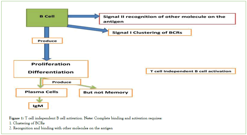
Figure 1: T cell independent B cell activation.
Note: Complete binding and activation requires:
1. Clustering of BCRs
2. Recognition and binding with other molecules on the antigen
T cell dependent B cell activation Th2
Antigen that triggers B cell activation with the help of T helper cells, protein antigens cannot cross link with multiple BCRs. Due to lack of repetitive and identical epitopes they require T cell activated it’s the four signal process.
Signal 1: Antigen peptide recognition and binding by B cells, and expressed stimulatory molecules CD40 and also activates T helper cell. B cells beside recognition also process and present them like APC.
Signal 2: Derived from B cell and T cell interactions, activates T helper cell binds to MHCII antigen peptide complex in the cell surface (depends of the antigen), and then CD40L expressed and bind CD40 molecule.
Signal 3: Provided by cytokines released by T helper cell stimulate B cell, The interactions between B cell and T helper cell induces the expression of new cytokines receptor under surface of B cell, T helper cell produces cytokine, these cytokine bind to the new cytokines receptor under surface of B cell as the result B cells start proliferate and differentiate, antibody secreting plasma cells and memory B cells (Figure 2).
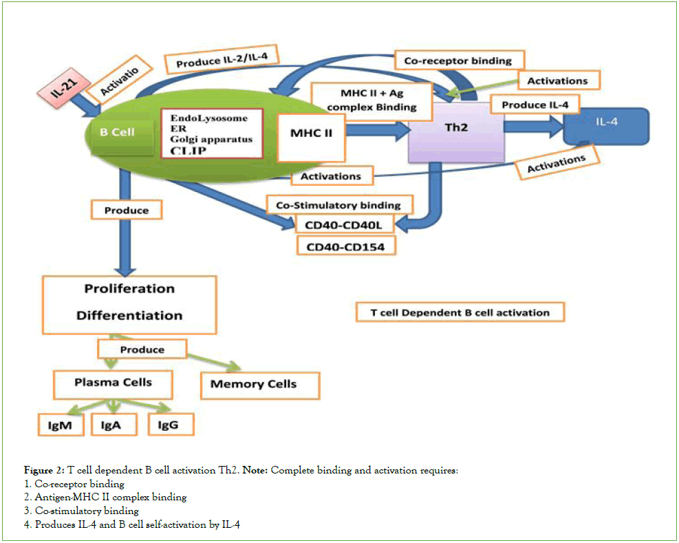
Figure 2: T cell dependent B cell activation Th2.
Note: Complete binding and activation requires:
1. Co-receptor binding
2. Antigen-MHC II complex binding
3. Co-stimulatory binding
4. Produces IL-4 and B cell self-activation by IL-4
T cell dependent B cell activation Tc1
Antigen that triggers B cell activation with the help of T helper cells, protein antigens cannot cross link with multiple BCRs. Due to lack of repetitive and identical epitopes, they require T cell activated, it’s the four signal process.
Signal 1: Antigen peptide recognition and binding by B cells, and expressed stimulatory molecules CD40 and also activates cytotoxic T cells 1. B cells beside recognition also process and present them like APC.
Signal 2: Derived from B cell and T cell interactions, activates cytotoxic T cells 1 binds to MHCI antigen peptide complex in the cell surface(depends of the antigen), and then CD40L expressed and bind CD40 molecule.
Signal 3: Provided by cytokines released by cytotoxic T cells 1 stimulate B cell, The interactions between B cell and cytotoxic T cells 1 induces the expression of new cytokines receptor under surface of B cell, cytotoxic T cells 1 produces cytokine, these cytokine bind to the new cytokines receptor under surface of B cell as the result B cells start proliferate and differentiate, antibody secreting plasma cells and memory B cells (Figure 3).
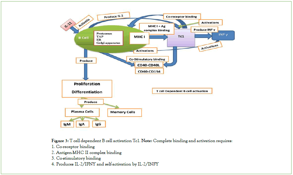
Figure 3: T cell dependent B cell activation Tc1.
Note: Complete binding and activation requires:
1. Co-receptor binding
2. Antigen-MHC II complex binding
3. Co-stimulatory binding
4. Produces IL-2/IFNY and self-activation by IL-2/INFY
T cell dependent macrophage activation
Antigen that triggers macrophage activation with the help of CD4 T cell, protein antigens cannot cross link with multiple receptors. Due to lack of repetitive and identical epitopes they require CD4 activated it’s the four signal process.
Signal 1: Antigen peptide recognition and binding by macrophage, and expressed stimulatory molecules CD80 and also activates CD4 T cell. Macrophage beside recognition also process and present them like APC.
Signal 2: Derived from macrophage and CD4 T cell interactions, activates CD4 T cell binds to MHCII antigen peptide complex in the cell surface and then CD80L expressed and bind CD28 molecule.
Signal 3: Provided by cytokines released by CD4 T cell and stimulate CD4 T cell. The interactions between macrophage and CD4 T cell induces the expression of new cytokines receptor under surface of CD4 T cell, CD4 T cell produces cytokine, these cytokine bind to the new cytokines receptor under surface of CD4 T cell as the result. Macrophage start proliferates and differentiate, antibody secreting effector CD4 T cell and memory CD4 T cells (Figure 4).
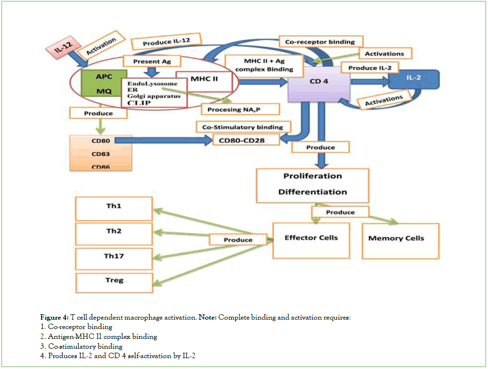
Figure 4: T cell dependent macrophage activation.
Note: Complete binding and activation requires:
1. Co-receptor binding
2. Antigen-MHC II complex binding
3. Co-stimulatory binding
4. Produces IL-2 and CD 4 self-activation by IL-2
T cell dependent dendritic cell activation
Antigen that triggers dendritic cell activation with the help of CD8 T cell, protein antigens cannot cross link with multiple receptors Due to lack of repetitive and identical epitopes they require CD8 activated it’s the four signal process.
Signal 1: Antigen peptide recognition and binding by dendritic cell, and expressed stimulatory molecules CD80L and also activates CD8 T cell.
Signal 2: Derived from dendritic cell and CD8 T cell interactions, activates CD8 T cell binds to MHC I antigen peptide complex in the cell surface and then CD80L expressed and bind CD28 molecule.
Signal 3: Provided by cytokines released by CD8 T cell and stimulate CD8 T cell. The interactions between dendritic cell and CD8 T cell induces the expression of new cytokines receptor under surface of CD8 T cell, CD8 T cell produces cytokine, these cytokine bind to the new cytokines receptor under surface of CD8 T cell as the result dendritic cell start proliferate and differentiate, antibody secreting effector CD8 T cell and memory CD8 T cells (Figure 5).
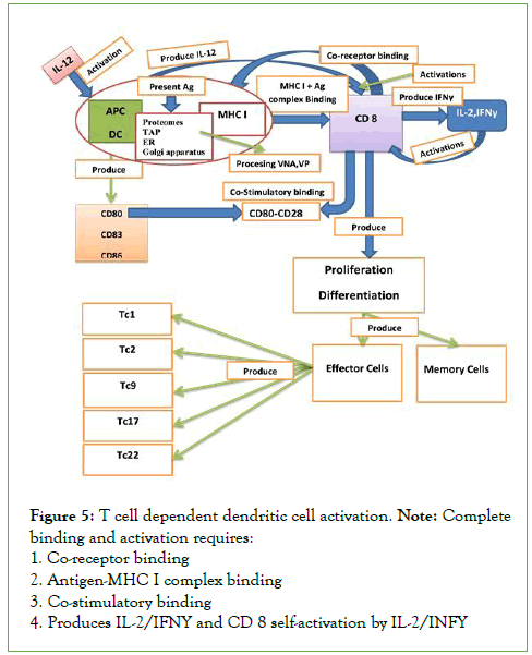
Figure 5: T cell dependent dendritic cell activation.
Note: Complete
binding and activation requires:
1. Co-receptor binding
2. Antigen-MHC I complex binding
3. Co-stimulatory binding
4. Produces IL-2/IFNY and CD 8 self-activation by IL-2/INFY
The Discovery Process: Innovation and Immunology
Efficacy refers how a vaccine performs under ideal lab conditions, such as those in a clinical trial. Effectiveness refers to how it performs in the real world. Vaccine AZEE CRA 01 from adjuvant, crucial for getting a stronger, longer-lasting, broader immune response especially among those with weakened immunity like pediatrics, children, pregnant women and elderly. Crucial for restore functions causes immune cells dysfunction and improve the normal immune function and balance modifications. Preventive and therapeutic and working with immunity to build protection.
When you get a vaccine AZEE CRA 01 it inhibits viral replication and spread humeral and cellular immune response, recognizes the invading virus the respiratory system, produces antibodies (IgM,IgG,IgA) and remembers the disease and how to fight it (entire normal signaling, effector and memory cell created).
Vaccine AZEE a safe and produce an immune response in the body, without causing illness, affecting immune cells function and with respect of biochemical properties of immune cells. Our immune systems are designed to remember, when we create suitable environment for natural immune cell signaling, mechanism and function. Once the immune cell kills foreign antigen, degraded, processed, present, produce the entire activation and naturally functioning to produce the specific antibodies for that specific antigen. We typically remain protected against a disease for years, decades or even a lifetime.
Zelalem Kiros Bitsue and his Vaccine Discoveries
1. SARS-COV-2 with all type of variants preventions and therapeutic.
2. Future respiratory infectious pandemic preventions.
3. Produce an immune response in the body, without causing illness, affecting immune cells function, and with respect of biochemical properties of Immune cells.
4. Without affecting genes transcriptions and expression.
5. Long lasting, very safe, well known components.
Zelalem Kiros Bitsue and his Immunologic Discoveries
His B cell discoveries
1. I discovered that B cell synthesis both intracellular and extracellular pathogen.
2. Further, B cell has proteasome, ER, GA and TAP and lysosome, endosome, GA and CLIP components.
3. In addition, B cell interacts both with cytotoxic T cell and T helper cells to make T dependent B cell activation.
His macrophages discoveries
4. I discovered that macrophages synthesis extracellular pathogen.
5. Further has lysosome, endosome, ER, GA and CLIP components.
6. In addition, macrophages interact with T helper cells to make CD4 T cell activation.
His dendritic cells discoveries
7. I discovered that dendritic cells synthesis intracellular pathogen.
8. Further has a proteasomes, ER, GA and TAP component.
9. In addition, dendritic cells interact with antigen complex peptide to make CD8 T cell activation.
Antigen that triggers B cell activation with the help of T helper cells and cytotoxic T cells 1, protein antigens cannot cross link with multiple BCRs. Due to lack of repetitive and identical epitopes, they require T cell activated it’s the four signal process (Figure 6).
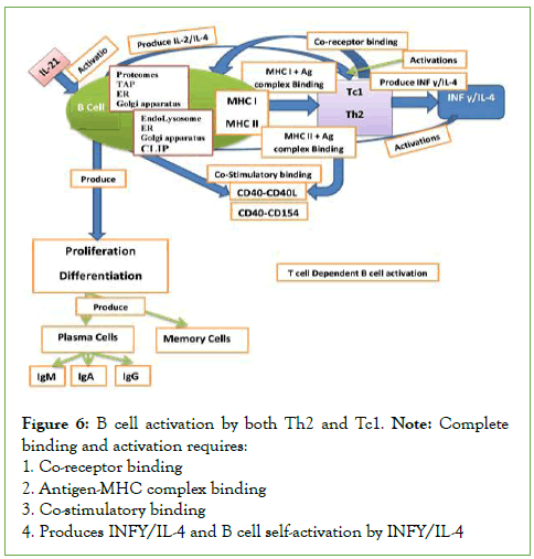
Figure 6: B cell activation by both Th2 and Tc1.
Note: Complete
binding and activation requires:
1.Co-receptor binding
2. Antigen-MHC complex binding
3. Co-stimulatory binding
4. Produces INFY/IL-4 and B cell self-activation by INFY/IL-4
Signal 1: Antigen peptide recognition and binding by B cells, and expressed stimulatory molecules CD40L and also activates T helper cells and cytotoxic T cells 1. B cells beside recognition also process and present them like APC.
Signal 2: Derived from B cell and T cell interactions, activates T helper cells and cytotoxic T cells 1 binds to MHC I or MHC II antigen peptide complex in the cell surface(depends of the antigen intracellular or extracellular), and then CD40L expressed and bind CD40 molecule.
Signal 3: Provided by cytokines released by T helper cells or cytotoxic T cells 1 stimulate B cell. The interactions between B cell and T helper cells or cytotoxic T cells 1 induces the expression of new cytokines receptor under surface of B cell, T helper cells or cytotoxic T cells 1 produces cytokine, these cytokine bind to the new cytokines receptor under surface of B cell as the result B cells start proliferate and Differentiate, antibody secreting Plasma cells and memory B cells.
In this case B cell synthesis both intracellular and extracellular pathogen. Further, B cell has proteomes, TAP, RER and Golgi Apparatus (GA). More so, B cell has Lysosome, Endosome. RER, Golgi Apparatus (GA) and CLIP components and also syntheses both MHC I and MHC II. In addition, B cell interacts both with cytotoxic T cell and T helper cells to make T dependent B cell activation.
Vaccine Azee Discovered and Mechanisms of Action 1 of 2
This is very important activations of B cells without the help of T cell. B cell also have TLR that recognized the inhalation molecules and then generate these second signals, after activation this B cells proliferate and differentiations in to plasma cells that only secretes IgM antibody but not memory cells production. In addition activated alveolar-macrophage, and the activated alveolar- macrophage start releasing IL-12 induces the innate immune defense to the respiratory pathogens (Figure 7).
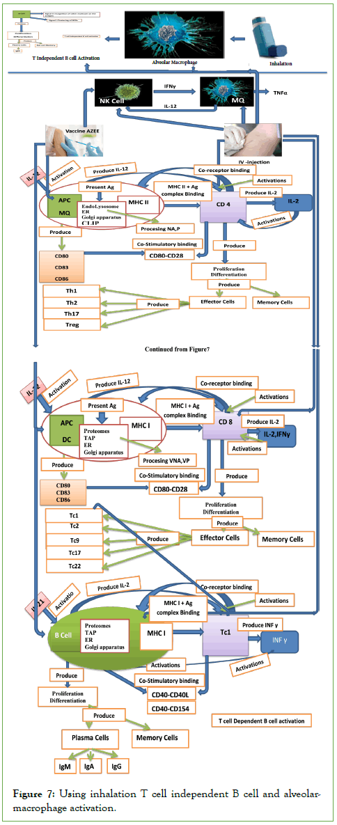
Figure 7: Using inhalation T cell independent B cell and alveolar-macrophage activation.
In addition, given IV-injection or oral tab depends on the patient conditions these crucial for restore functions causes immune cells dysfunction and improve the normal immune function and balance modifications, both in innate and adaptive immune cells (intracellular and extracellular). Further, given vaccine AZEE for activation of NK cell, macrophages, dendritic cells and B cells it induces both the innate and adaptive immune activation, the entire signaling and mechanisms restores. The inhalation and IV- injection given enough time to the immune cells, and the virus not to escape from the immune cells, more so, the virus even escapes the number of the replicated virus to become lower in number and incapables of creating cytokine storms. Vaccine AZEE then induces immune cells to do the normal immune signaling and mechanisms such us digesting, processing, presenting, synthesizing, and then secreting/producing specific antibodies for that specific antigen without causing illness, affecting immune cells function and with respect of biochemical properties of immune cells.
Conclusion
Inflammatory transcription factors such as NF-B mediate the activation and overexpression of several inflammatory cytokines. Furthermore, a recent study found that the AT1R phosphorylates JAK2 in lung cells, activating STAT-3 transduction to the nucleus. STAT-3 is a signal transducer and transcription activator that initiates the active transcription of inflammatory cytokines. AZEE CRA 01 vaccine with adjuvant is critical for eliciting a stronger, longer-lasting, broader immune response, particularly in those with weakened immunity important for restoring immune cell function and improving normal immune function, balance modifications and the status of vaccine AZEE CRA 01 is under development (Safety and Efficacy test-USA).
References
- Bestle D, Heindl MR, Limburg H, Pilgram O, Moulton H, Stein DA, et al. TMPRSS2 and furin are both essential for proteolytic activation of SARS-CoV-2 in human airway cells. Life Sci Alliance. 2020;3(9):e202000786.
[Crossref] [Google Scholar] [PubMed]
- Bhattacharya M, Sharma AR, Mallick B, Sharma G, Lee SS, Chakraborty C. Immunoinformatics approach to understand molecular interaction between multi-epitopic regions of SARS-CoV-2 spike-protein with TLR4/MD-2 complex. Infect Genet Evol. 2020;85:104587.
[Crossref] [Google Scholar] [PubMed]
- Bobati SS, Naik KR. Therapeutic plasma exchange-an emerging treatment modality in patients with neurologic and non-neurologic diseases. J Clin Diagn Res. 2017;11(8):EC35-EC37.
[Crossref] [Google Scholar] [PubMed]
- Fürsch J, Kammer KM, Kreft SG, Beck M, Stengel F. Proteome-wide structural probing of low-abundant protein interactions by cross-linking mass spectrometry. Anal Chem. 2020;92(5):4016-4022.
[Crossref] [Google Scholar] [PubMed]
- Henderson LA, Canna SW, Schulert GS, Volpi S, Lee PY, Kernan KF, et al. On the alert for cytokine storm: immunopathology in COVID‐19. Arthritis Rheumatol. 2020;72(7):1059-1063.
[Crossref] [Google Scholar] [PubMed]
- Kanimozhi G, Pradhapsingh B, Singh Pawar C, Khan HA, Alrokayan SH, Prasad NR. SARS-CoV-2: pathogenesis, molecular targets and experimental models. Front Pharmacol. 2021;12:638334.
[Crossref] [Google Scholar] [PubMed]
- Kaur T, Kapila S, Kapila R, Kumar S, Upadhyay D, Kaur M, et al. Tmprss2 specific miRNAs as promising regulators for SARS-CoV-2 entry checkpoint. Virus Res. 2021;294:198275.
[Crossref] [Google Scholar] [PubMed]
- Kuba K, Imai Y, Ohto-Nakanishi T, Penninger JM. Trilogy of ACE2: A peptidase in the renin–angiotensin system, a SARS receptor, and a partner for amino acid transporters. Pharmacol Ther. 2010;128(1):119-128.
[Crossref] [Google Scholar] [PubMed]
- Li F. Structure, function, and evolution of coronavirus spike proteins. Annu Rev of Virol. 2016;3(1):237-261.
[Crossref] [Google Scholar] [PubMed]
- Li F, Li W, Farzan M, Harrison SC. Structure of SARS coronavirus spike receptor-binding domain complexed with receptor. Science. 2005;309(5742):1864-1868.
[Crossref] [Google Scholar] [PubMed]
- Lima MA, Skidmore M, Khanim F, Richardson A. Development of a nano-luciferase based assay to measure the binding of SARS-CoV-2 spike receptor binding domain to ACE-2. Biochem Biophys Res Commun. 2021;534:485-490.
[Crossref] [Google Scholar] [PubMed]
- Mehta P, McAuley DF, Brown M, Sanchez E, Tattersall RS, Manson JJ. COVID-19: consider cytokine storm syndromes and immunosuppression. Lancet. 2020;395(10229):1033-1034.
[Crossref] [Google Scholar] [PubMed]
- Qi J, Zhou Y, Hua J, Zhang L, Bian J, Liu B, et al. The scRNA-seq expression profiling of the receptor ACE2 and the cellular protease TMPRSS2 reveals human organs susceptible to SARS-CoV-2 infection. Int J Environ Res Public Health. 2021;18(1):284.
[Crossref] [Google Scholar] [PubMed]
- Sanda M, Morrison L, Goldman R. N-and O-glycosylation of the SARS-CoV-2 spike protein. Anal Chem. 2021;93(4):2003-2009.
[Crossref] [Google Scholar] [PubMed]
- Seif F, Aazami H, Khoshmirsafa M, Kamali M, Mohsenzadegan M, Pornour M, et al. JAK inhibition as a new treatment strategy for patients with COVID-19. Int Arch Allergy Immunol. 2020;181(6):467-475.
[Crossref] [Google Scholar] [PubMed]
- Sriram K, Insel PA. A hypothesis for pathobiology and treatment of COVID‐19: The centrality of ACE1/ACE2 imbalance. British journal of pharmacology. 2020;177(21):4825-4844.
[Crossref] [Google Scholar] [PubMed]
- Tikellis C, Thomas MC. Angiotensin-converting enzyme 2 (ACE2) is a key modulator of the renin angiotensin system in health and disease. Int J Pept. 2012;2012:256294.
[Crossref] [Google Scholar] [PubMed]
- Vankadari N, Wilce JA. Emerging COVID-19 coronavirus: glycan shield and structure prediction of spike glycoprotein and its interaction with human CD26. Emerg Microbes Infect. 2020;9(1):601-604.
[Crossref] [Google Scholar] [PubMed]
- Wan Y, Shang J, Graham R, Baric RS, Li F. Receptor recognition by the novel coronavirus from Wuhan: an analysis based on decade-long structural studies of SARS coronavirus. J Virol. 2020;94(7):e00127-20.
[Crossref] [Google Scholar] [PubMed]
- Yan R, Zhang Y, Li Y, Xia L, Guo Y, Zhou Q. Structural basis for the recognition of SARS-CoV-2 by full-length human ACE2. Science. 2020;367(6485):1444-1448.
[Crossref] [Google Scholar] [PubMed]
- Ye Q, Wang B, Mao J. The pathogenesis and treatment of the Cytokine Storm ‘in COVID-19. J Infect. 2020;80(6):607-613.
[Crossref] [Google Scholar] [PubMed]
- Zang R, Castro MF, McCune BT, Zeng Q, Rothlauf PW, Sonnek NM, et al. TMPRSS2 and TMPRSS4 promote SARS-CoV-2 infection of human small intestinal enterocytes. Sci Immunol. 2020;5(47):eabc3582.
[Crossref] [Google Scholar] [PubMed]
- Zheng YY, Ma YT, Zhang JY, Xie X. COVID-19 and the cardiovascular system. Nat Rev Cardiol. 2020;17(5):259-260.
Citation: Bitsue ZK (2023) Impact of Immunologic Discoveries on the Vaccine. J Vaccines Vaccin. 14:515.
Copyright: © 2023 Bitsue ZK. This is an open-access article distributed under the terms of the Creative Commons Attribution License, which permits unrestricted use, distribution, and reproduction in any medium, provided the original author and source are credited.

