Indexed In
- Open J Gate
- Genamics JournalSeek
- Academic Keys
- JournalTOCs
- ResearchBible
- China National Knowledge Infrastructure (CNKI)
- Scimago
- Ulrich's Periodicals Directory
- Electronic Journals Library
- RefSeek
- Hamdard University
- EBSCO A-Z
- OCLC- WorldCat
- SWB online catalog
- Virtual Library of Biology (vifabio)
- Publons
- MIAR
- Scientific Indexing Services (SIS)
- Euro Pub
- Google Scholar
Useful Links
Share This Page
Journal Flyer

Open Access Journals
- Agri and Aquaculture
- Biochemistry
- Bioinformatics & Systems Biology
- Business & Management
- Chemistry
- Clinical Sciences
- Engineering
- Food & Nutrition
- General Science
- Genetics & Molecular Biology
- Immunology & Microbiology
- Medical Sciences
- Neuroscience & Psychology
- Nursing & Health Care
- Pharmaceutical Sciences
Research - (2023) Volume 14, Issue 6
Expression of some genes involved in epigenetic in breast cancer cell line (MCF7): The effect of gold nanoparticles
Mojgan Noroozi-Karimabad1*, Marzie Salandari Rabori1, Omid Azizian-Shermeh2 and Seyedeh Atekeh Torabizadeh32Department of Chemistry, Faculty of Science, University of Sistan and Baluchestan, Zahedan, Iran
3Pharmaceutics Research Center, Institute of Neuropharmacology, Kerman University of Medical Sciences, Kerman, Iran
Received: 01-Nov-2023, Manuscript No. jnmnt-23-23210; Editor assigned: 03-Nov-2023, Pre QC No. jnmnt-23-23210(PQ); Reviewed: 17-Nov-2023, QC No. jnmnt-23-23210; Revised: 24-Nov-2023, Manuscript No. jnmnt-23-23210(R); Published: 30-Nov-2023, DOI: 10.35248/2157-7439.23.14.695
Abstract
Background: Nano medicine is defined as the utilizing nanoparticles for treatment and diagnosis of diseases. Gold nanoparticles to be surveyed. Green synthesis is another alternative as an economically viable, simple, and environmentally friendly method for synthesizing gold nanoparticles. Here we have investigated the potential of gold nanoparticles synthesis in inhibiting the expression of self-renewal regulatory factors and cancer stem cell gene and apoptosis genes in a leukemia cell line MCF7.
Methods: Using green chemistry technique, gold nanoparticles were synthesized using walnut green external shell, where the green shell of walnut is a reducer and stabilizer for providing gold nanoparticles. These nanoparticles were examined using x-ray diffraction (XRD) and transmission electron microscopy (TEM) to determine their size, structure, and shape. Cell proliferation inhibition was tested using MTT assay. Real-time-PCR method was applied to indicate the fold changes of P53, NANOG, SOX, OCT4, and CAS expression against β-actin. Data were analyzed by Two-Way ANOVA Differences were considered significant if (P<0.05).
Result: A significant variation was observed in cell viability when different gold nanoparticles’ concentrations were applied for 24 h compared to control with regard to the cellular viability (P <0.05). Real time-PCR analysis showed that the NANOG, OCT4, and SOX gene expression levels down-regulated while CAS and P53 were up-regulated compared to untreated control cells and cells treated (P<0.05).
Conclusion: The results revealed that gold nanoparticles may hinder the proliferation of cancer in in vitro, resulting in the development of a desired treatment agent for treating MCF7 cancer. To sum up, the current study offers promising information on stem-cell genes expression in MCF7 cells.
Keywords
Gene; Antitumor; Gold nanoparticles; MCF7; Self-renewal; Apoptosis
INTRODUCTION
The number of patients with cancer is rising. Thus, it is critical to find influential ways to overcome these pathogens and cancers. Nanoscale m
aterials have attracted the attention of scholars and materials scientists. The particle size reduction from micro to nano has contributed to the appearance of specific features including high electrical conductivity, hardness, high reactivity, increased surfaceto- volume ratio, and altered biological activit [1], which make the particles interact more meritoriously with bacterial membranes, microbes, and cancerous cells. So, they discharge metal ions along the membrane [2]. The toxicity of metal NPs, such as copper, gold, zinc, and titanium, has been notably considered for therapeutic alternatives [3,4]. Gold, copper, and silver nanoparticles have been of great interest as they possess intrinsic anti-cancer, antibacterial, and antioxidant features due to their convenient and speedy usage, low-concentration toxicity, availability, and lack of environmental risks [5]. Gold NPs, with various morphological structures, may own multifunction biological activities like anti-cancer and antibacterial roles. Studies have also argued that the impact of nanoparticles relies on size [6]. However, there are few studies on the professional performance of NPs and toxicology. Gold NPs are NPs used in medicine, catalysts, agriculture, biology, covering, electronic devices, and drug delivery, etc. Gold NPs are going to be used for cancer treatment and are evaluated as medicine carriers, contrast agents, photothermal agents, and radio sensitizers. Gold NPs functionalized stably with covalently attached oligonucleotides activate immune-related genes and pathways in peripheral blood mononuclear cells of human, but not an immortalized, lineagerestricted cell line. Simple biocompatible processes are required for non-toxic NPs’ low-cost synthesis. Fungi, bacteria, yeasts, actinomycetes, and plants have been applied in the biological procedures as reducing factors for the synthesizing different NPs [7]. The NPs biogenic synthesis from plant extract is possibly an influential technique to raise innocuous, facile, and biocompatible NPs. Some plant extracts, like, Epigallocatechin gallate, Tamarindus indica, Citrus paradise Calotropis procera Murraya koenigii and Hibiscus sabdariffa [8] have been employed as reducing agents for the synthesis of NPs. Muthukumar et al. found that the extracts of A. spinosus aqueous leaf enjoy therapeutic features and rich phytochemical components having great antioxidant capacity and serving as an reducing agent for the synthesis of NPs. Elumalai and Velmurugan explained the green synthesis of ZnO NPs by A. The synthesized gold NPs of walnut green shell were applied for the treatment of breast cancer. Cancer stem cells are similar to stem cells in numerous aspects like exhibiting immortality, self-renewal, and differentiation. The cancer stem cells are associated with the fact that only some cancer cells show the special capability of selfrenewal followed by infinite proliferation. Nevertheless, these cells’ descriptions are limited because of their small number and not having suitable markers. The OCT-4 and NANOG, SOX2 serving as pluripotent-asocial agents are the main self-renewal regulators in embryonic and cause pluripotent traits of stem cells. Apoptosis induction is a possible desired procedure to prevent cancer or treat furring different steps. Therefore, the induction of apoptosis using these plants is associated [9].
With a great technique to put an end to the progression and development of carcinogenesis besides eleminating genetically injured, reinitiated, or neoplastic cells. CAS and P53 have been assumed the main apoptosis regulators in cancer cells [10]. In the current work, the impact of potential of gold NPs synthesis was developed and evaluated on MCF7 cell through the assessment of the expression and evaluation of the balancing NANOG, SOX, OCT4, P53, And CAS genes in mRNA and protein levels to the potential mechanism of molecules that may be involved.
METHODS AND EXPERIMENTS
Synthesis of gold NPs
The A. spinosus hydro-alcoholic leaf extraction was done by a soxhlet apparatus. The extracts were kept at 5 °C in a container that was airtight.
For gold NPs synthesis, 1 mM concentration of HAuCl4.3H2O was prepared, and 4 ml of it was added to 2 ml extract of walnut at ambient temperature. In fact, the extract of walnut was added as a reducing agent. The pH was 5.4. Color solution change from green to purple depicts synthesized gold NPs.
Optimum conditions for producing bigger gold NPs
To make high-quality and big gold NPs, some factors were altered such as temperature, extract volume, pH, and gold salt concentration.
Cell culture and treatment
MCF7 cells was prepared from the National Cell Bank of Iran (NCBI, Tehran, Iran). Cell culture was done in a 25-cm2 flask at RPMI 1960 supplement with 10% FBS, 1% glutamine plus 100 IU/ ml penicillin, and 100 IU/ml streptomycin, which was incubated at 37°C in 5% CO2 (7).
Measurement of cell viability/proliferation by MTT assay
MTT assay was used to assess MCF7 cells’ viability or proliferation of after being exposed to gold NPs. MCF7 cancer cells were put in 96-well plates in 200 μl regular medium nightlong long. Afterwards, they were treated with differing concentrations for 24 h. This method was previously presented 20 μl of MTT solution (5 mg/ml in PBS) was poured into every well and incubated for 4 h at a cell-culture incubator. The supernatant was removed, and 100 μl DMSO was added to every well. Then, optical density (OD) was obtained by an ELISA microplate reader at 570 nm. The viability or proliferation difference between the treated cells and the control was measured using the equation below:
Viability rate (%): (OD of the treated cells/ OD of the control cells) × 100(7).
RNA ISOLATION, CDNA SYNTHESIZE AND REAL-TIME PCR
RNA extraction
Total RNA was separated from the MCF7 cells by RNeasy Mini Kit (QIAGEN, Hilden, Germany) one-step method according to the manufacturer’s guidelines (Bioneer, Korea), accompanied by treatment with different Gold NPs
Concentrations for 24 h.The samples of RNA were dissolved in diethylpyrocarbonate (DEPC)-treated water.The RNA values were indicated using a PD-303UV spectrophotometer (APEL, Japan). The isolated RNA fidelity was examined by electrophoresis on 1% agarose and staining with DNA Green Viewer™. Three bands with the Gel Doc were observed from every sample of RNA.
DNA synthesis
Employing a reverse transcriptase kit (Bioneer, Korea), complementary DNA (cDNA) was made up. The obtained cDNA was either kept at -20 ºC or rapidly applied for quantitative RT-PCR. Then, the RNA converted to cDNA through reverse transcriptase enzyme was employed as a RT-PCR technique target.
RT-PCR
The gene expression of NANOG, OCT4, SOX, P53 and CAS were detected, employing RT PCR. To approach this, the synthesized cDNA was taken at this step and was gently mixed with master mix contained DNA Taq polymerase. It contains DNA Taq polymerase mixture and allowing RT-PCR run for 40 cycles, as shown in Table-1 Relative quantification of gene expression was performed in triplicate. The mRNA expression was determined using SYBR Premix Ex Taq™ kit (Takara Bio, USA). The primers sequences were demonstrated in Table 1. The β-actin was utilized as a reference gene, and the target genes’ expression is associated with its expression. Relative expression was measured by the comparative 2-ΔΔCt equation.
| Gene | Forward (5'â??3') | Reverse (5'â??3') |
|---|---|---|
| Oct-04 | CCTGAAGCAGAAGAGGATCACC | AAAGCGGCAGATGGTCGTTTGG |
| SOX2 | GCTACAGCATGATGCAGGACCA | TCTGCGAGCTGGTCATGGAGTT |
| NANOG | CTCCAACATCCTGAACCTCAGC | CGTCACACCATTGCTATTCTTCG |
| p53 | TGAAGCTCCCAGAATGCCAG | GCTGCCCTGGTAGGTTTTCT |
| Caspase-3 | CTCGGTCTGGTACAGATGTCGATG | GGTTAACCCGGGTAAGAATGTGCA |
| β-actin | CACACCTTCTACAATGAGC | ATAGCACAGCCTGGATAG |
Table 1: The sequences of the employed primers in this study
Instrumental analysis
Prism and SPSS software (version 21) were used to analyze the data statistically. All experiments, in every individual sample were executed in triplicates and all attained consequences, as the mean ± SEM were indicated for those three. Data were analyzed by Two- Way ANOVA Differences were considered significant if (P<0.05)
RESULTS
Structural analysis of synthesized NP
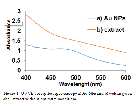
To study the structural analysis of synthesized nanoparticles, the UV-Vis spectroscopy was detected at 200-800 nm wavelength range. A broad peak was observed at 480 nm (Figure 1).
Figure 1: UV-Vis absorption spectroscopy a) Au NPs and b) walnut green shell extract without optimum conditions
Results of optimum pH and volume and concentration and temperature
The comparison of these solutions’ spectrum indicated that pH=6 solution has maximum concentration. Therefore, the optimum pH = 6. Various solutions’ absorption spectrum reveals that more NPs of gold was made in 2 ml essence. So, 2 ml was an optimum volume for the extract. Five differing concentrations of gold salt were obtained. The highest absorption is associated with gold salt with 3 mM concentration. Moreover, solutions with the so-called desired conditions were provided and studied at different temperatures. It shows more adsorption spectrum at 55°C; hence, 55°C is the optimum rate.
Characterization of the gold nps by xrd and tem
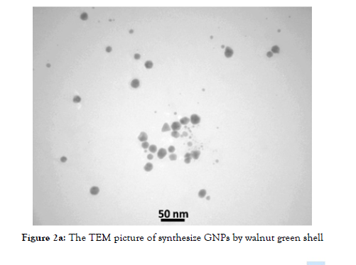
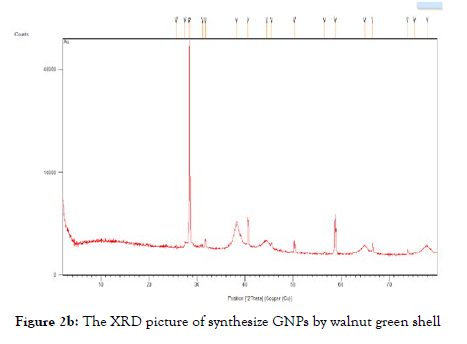
To estimate the NPs synthesis size and assess their shape, TEM was employed, confirming that 10-50 nm spherical and triangular gold NPs were synthesized. To indicate crystalline NPs phase and provide information on unit cell dimensions, XRD was used that showed successful NPs synthesis. (Figures 2a and 2b) show the results of TEM and XRD, respectively.
Figure 2a: he TEM picture of synthesize GNPs by walnut green shell
Figure 2b: The XRD picture of synthesize GNPs by walnut green shell
RESULTS OF THE ANTI-CANCER POTENTIAL OF GOLD NPS
Concentration-dependent of gold NPs on cancer cell viability/ proliferation
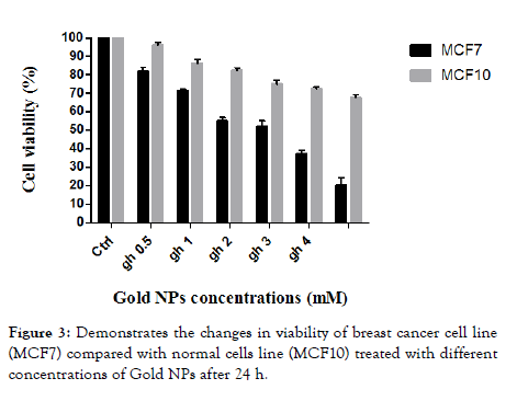
MTT assay evaluated the impact concentrations of Gold NPs on the viability or proliferation of breast cancer cell line (MCF7) compared with normal cells line (MCF10). The rise in concentration has decreased cancer cells’ viability compared with normal cells (Figure 3). The Table 2 Exhibited the IC50 values (mM) of Gold NPs against MCF7 and MCF10 Cells after 24 h of treatment.
Figure 3: Demonstrates the changes in viability of breast cancer cell line (MCF7) compared with normal cells line (MCF10) treated with different concentrations of Gold NPs after 24 h.
Gold NPs altered CAS and P53 mRNA levels on expression apoptosis gene: The mRNA expression of P53 and CAS rose meaningfully in MCF7 cells that were treated with Gold NPs in comparison with the control cells after 24 h. Values are the average of triple determinations with the SD. indicated by error bars (P< 0.05).
* = significant in compared with Con group (P < 0.05).
Gold NPs altered NANOG, OCT4 and SOX mRNA levels on expression of self-renewal regulatory factors and cancer stem cell gene.
To further corroborate the results of this study, the expression levels of NANOG, OCT4 and SOX mRNA were analyzed by realtime- PCR in MCF7 cells treated with Gold NPs in comparison with the control cells (untreated) after 24, 48 and 72 h under IC50 concentrations. . The results of real-time-PCR analysis showed that NANOG, OCT4 and SOX mRNA expression levels were decreased in NB4 cells treated with Gold NPs in comparison with the control cells (untreated) cells after 24 incubation.
Values are the average of triple determinations with the SD. indicated by error bars (P< 0.05).
*Significant in compared with Con group (P < 0.05).
DISCUSSION
Although there are decent advancements regarding novel medicine and pharmaceutical biotechnology, cancer is still a main cause of death in the globe. Thus, it is necessary to find influential procedures to handle cancers. Metal NPs are novel ways to confront cancers. The most common one among women is breast cancer. Since it has a widespread effect, it is assumed a critical public health issue requiring further nanotechnology and molecular studies to settle prognosis and treatment possibilities. Compounds extracted from plants and nanomaterials are considered as a desired and practical option for MCF-7 therapy and other cells of cancer. The toxicity of these anticancer compounds is lower and chemo-preventive, which make them a focus of cancer-related studies. However, they have limited capability to fight a cancer site because of insufficient solubility, bioavailability and structural deformation. Gold NPs have recently been the focus of scholars due to their sophisticated medicinal uses. In the present study, investigation on the biological effects of Au NPs on MCF7 cancers. Our results demonstrated that nanoparticles with increased concentration led to decreased survival, increased apoptosis genes expression such as CAS and P53 in MCF7 cancer cells. And also statistical analysis of our real time-PCR data showed that the mRNA expression of NANOG, OCT4 and SOX was significantly decreased in MCF7 cells treated with Gold NPs compared to control cells after 24 h. Overall, our results provide solid evidence to back up previous research. Qi Chen concluded that the SPARC expression declined after being administered with Au NPs based on a certain dose. It has been demonstrated that albumin can be transferred into the tumor by a SPARC-mediated transport mechanism. Reduced expression of SPARC protein manifests that less albumin is there for the development of tumor, which is possibly another cause of why S4A–BSA–Au NPs can increase the anticancer influence of S4A. Accordingly, the drug delivery system B-BANC-NPs proposes a novel method to pharmaceutically improve hydrophobic natural medicine. Zhao et al. proved that the combined therapy of cancer cells utilizing Fe3O4@MTX-LDH/Au for synergistic hyperthermia ablation and chemotherapy shows greater treatment effectiveness compared to only one treatment, underlining the platform potential for the treatment of cancer. Shengtao Wang carried out in situ synthesis of super organism-like Gold NPs in micro gels with ultra-wide absorption in visible and near-infrared areas for combined treatment of cancer, confirming that micro gels and nano composites may load concurrently photosensitive drugs due to promising loading capability, and may be employed for combined photo thermal and photodynamic therapies. Xinjie Wang et al. synthesized magnetic cores (Fe3O4) by a fabrication technique that involved Fe2+ and Fe3+ coprecipitation. Afterwards, the magnetic NPs were encapsulated by layers of chitosan. Then, magnetite-gold composite NPs were synthesized with spherical shapes and sizes of 20-30 nm, applying trisodium citrate as a natural reduction agent. The most promising cytotoxicity outcomes of Fe3O4@CS/ Au NPs were detected regarding the HCT 116 cell line. The current NPs may be employed for treating many gastro-duodenal cancers particularly gastric, colon, and pancreatic cancers. Al-Radadi found that seed and pulp oil extract mediated gold NPs demonstrated promising outcomes on anti-inflammatory, anti-diabetic, anticancer and anti-Alzheimer features. HepG2, HCT-116, and MCF-7 cell lines were discarded by the cytotoxic influences of the NPs and extracts. The results of the study on gold NPs generated from seed and pulp oil extract proved that they can be utilized as vectors in biomedical usages like gene delivery, medication administration, or as biosensors where they face blood. Biogenic Gold NPs seed oil show potential anticancer influences on colon cancer cells. The total elimination, absorption, and anti-tumor features of the test materials are affected by surface chemistry, physical properties, and dosage dependency. Metallic NPs damage DNA, causing the death of malignant cells though the precise mechanism of NPs cytotoxicity is not still known. The functional group interaction of cellular proteins with NPs, leading to DNA injury, was explained as Ag and Au NPs cytotoxicity method. Au-NPs have also shown to induce oxidative stress via generation of reactive oxygen species (ROS), causing DNA damage and killing cancer cells. Overall, gold NPs can be applied in the biomedical activities. Nevertheless, invivo studies have to be developed to assess the precise cytotoxicity of the bio inspired NPs. The impact of metal NPs on various cancer cell lines is different, emphasizing different cancer cell lines’ resistance against these NPs. The biomolecules encapsulated on the surface have plenty of hydroxyl groups and develop complexes with metal ions. Therefore, reduced toxicity is observed when MCF-7 cells uptake and metabolize NPs. The Au NPs’ IC50 values were 3 mM. Metal atoms offer a kinetic restriction for releasing metal into solution, reducing the rate of dissolution and decreasing cytotoxicity. But, higher NPs concentrations for 24 h brought about significant synthesized NPs, showing less cytotoxicity and MCF cell reduction. Also, higher free ions’ concentration can bring about homeostasis imbalance and aberrant cellular responses, such as epigenetic events, DNA damage, cytotoxicity, oxidative stress, and inflammatory processes.
Furthermore, the sphere-forming ability of cancer cells was impaired upon Gold NPs treatment, proposing that Gold NPs might have been interfered with the tumor-initiating ability of these cells. Although previous reports have proposed an evident link between multipotency- and pluripotency-associated marker expression and the sphere-forming ability of cancer cells, the mechanism of the decrease in sphere-forming ability in MCF7 cells upon Gold NPs treatment deserves to be further clarified. An emerging body of evidences reported that a subpopulation of a tumor carries a distinct molecular gene and is selectively resistant to chemotherapy. In recent years, many therapeutic methods have been developed to identify and specifically target these subpopulations in different tumor tissues. Increases in the expression of stem-ness markers and gene, including NANOG, OCT were shown to play a critical role in chemoresistance to cisplatin, doxorubicin, docetaxel, 5-fluorouracil and carboplatin. These therapies mostly concerned the inhibiting of self-renewal of cancer stem cells, inducing their differentiation. However, therapy strategies with existing anti-cancer agents like cisplatin, oxaliplatin and 5-fluorouracil shown to enhance cell viability and promote the self-renewal and survival of cancer stem cells in different cancer models. Therefore, approaching novel agents with capacity suppress the stem-like features of cancer cells and simultaneously inhibiting their proliferation is of importance. Considerable levels of efforts have been made toward replacing anticancer therapy agents with compounds to eradicate the undesirable effects of the chemicals during treatment and Gold NPs successfully inhibited pluripotency-, multipotency- and angiogenesis-associated genes like NANOG, OCT-4. To study the underlying mechanism by which Gold NPs enhances the apoptosis in NB4, we performed some experiments on the expression of genes expression involved in the apoptosis pathway. The decreased apoptosis is a critical step in the procedure of cancer evolution it was well established that the mitochondrial apoptosis pathway is the most significant pathway by regulating the ratio of apoptotic proteins Bcl-2 and Bax evidences also shown that Bcl-2 suppresses apoptosis by blocking of the release of cytochrome C from mitochondria and further initiation of apoptosis while Bax has an apoptosis-promoting effect by antagonizing Bcl-2. The balance between these two apoptosis regulatory genes, (the Bcl-2 and Bax) is a key phenomenon in cell survival and death since the increase in Bax/Bcl-2 ratios contributes to the efflux of cytochrome C from mitochondria and activates the intrinsic mitochondrial apoptotic pathway. Consequently, the changes in Bax and Bcl-2 expression determines tendency of apoptosis in cells. Increased Bax/Bcl-2 lead to activate caspase-3 activation and subsequently and result in cell death. It is well demonstrated that apoptosis plays fundamental parts in response to chemotherapy, recommending a relationship between drug-induced apoptosis and therapeutic efficiency in leukemic patients. As a result, the analysis of mitochondrial apoptotic genes may lead to increment of a key prognostic tool to predict resistance to chemotherapy compounds. Independently, the functions of Bcl-2 and Bax have been widely studied in prostate and other cancers, providing facts of their opposing function in cell survival and apoptosis.
CONCLUSION
The Gold NPs are potential anticancer agents and may cure metastatic cancers like breast cancer. The current study offers that Au NPs can be synthesized for stability and penetrate cancer cells. Thus, small Au NPs can be applied as an anti-cancer agent in pharmaceutical biotechnology. Since NPs can induce some problems, it should be evaluated in animal models regarding vivo side effects. Moreover, the present argues that Au NPs may confront metastatic cancer cells. A novel strategy is proposed for to pharmaceutically improve hydrophobic natural medicines.
INFORMED CONSENT
No informed consent
ACKNOWLEDGEMENTS
This project was supported by a grant from the Rafsanjan University of Medical Sciences.
REFERENCES
- Ilinskaya AN, Shah A, Enciso AE, Chan KC, Kaczmarczyk JA. Nanoparticle physicochemical properties determine the activation of intracellular complement. Nanomedicine: Nanotechnology, Biology and Medicine. 2019; 17: 266-75.
- Yang G, Xie J, Hong F, Cao Z, Yang X. Antimicrobial activity of silver nanoparticle impregnated bacterial cellulose membrane: Effect of fermentation carbon sources of bacterial cellulose. Carbohydrate Polymers. 2012; 87(1): 839-45.
- Babaei S, Bajelani F, Mansourizaveleh O, Abbasi A, Oubari F. A study of the bactericidal effect of copper oxide nanoparticles on Shigella sonnei and Salmonella typhimurium. Journal of Babol University of Medical Sciences. 2017; 19(11): 76-81.
- Abbasi A, Ghorban K, Nojoomi F, Dadmanesh M. Smaller copper oxide nanoparticles have more biological affects versus breast cancer and nosocomial infections bacteria. APJCP. 2021; 22(3): 893.
- Kukia NR, Rasmi Y, Abbasi A, Koshoridze N, Shirpoor A. Bio-effects of TiO2 nanoparticles on human colorectal cancer and umbilical vein endothelial cell lines. APJCP. 2018; 19(10): 2821.
- Ali K, Saquib Q, Ahmed B, Siddiqui MA. Bio-functionalized CuO nanoparticles induced apoptotic activities in human breast carcinoma cells and toxicity against Aspergillus flavus: an in vitro approach. Process Biochemistry. 2020; 91: 387-97.
- Salandari Rabori M, Karimabad MN, Hajizadeh MR. Facile, low-cost and rapid phytosynthesis of stable and eco-friendly gold nanoparticles using green walnut shell and study of their anticancer potential. World Cancer Res J. 2021; 8: 1-8.
- Qian X, Peng XH, Ansari DO, Yin-Goen Q. In vivo tumor targeting and spectroscopic detection with surface-enhanced Raman nanoparticle tags. Nature biotechnology. 2008; 26(1): 83-90.
- Koupaei MH, Shareghi B, Saboury AA, Davar F, Semnani A, Evini M. Green synthesis of zinc oxide nanoparticles and their effect on the stability and activity of proteinase K. RSC advances. 2016; 6(48): 42313-23.
- Jayaseelan C, Rahuman AA, Kirthi AV, Marimuthu S. Novel microbial route to synthesize ZnO nanoparticles using Aeromonas hydrophila and their activity against pathogenic bacteria and fungi. Spectrochimica Acta Part A: Molecular and Biomolecular Spectroscopy. 2012; 90: 78-84.
Indexed at, Google Scholar, Crossref
Indexed at, Google Scholar, Crossref
Indexed at, Google Scholar, Crossref
Indexed at, Google Scholar, Crossref
Indexed at, Google Scholar, Crossref
Indexed at, Google Scholar, Crossref
Citation: Noroozi-Karimabad M, Rabori MS, Azizian-Shermeh O, Torabizadeh SA (2023) Expression of Some Genes Involved in Epigenetic in Breast Cancer Cell line (MCF7): The effect of Gold Nanoparticles. J Nanomed Nanotech. 14: 695.
Copyright: ©2023 Noroozi-Karimabad M, et al. This is an open-access article distributed under the terms of the Creative Commons Attribution License, which permits unrestricted use, distribution, and reproduction in any medium, provided the original author and source are credited.


