


Indexed In
- Online Access to Research in the Environment (OARE)
- Open J Gate
- Genamics JournalSeek
- JournalTOCs
- Scimago
- Ulrich's Periodicals Directory
- Access to Global Online Research in Agriculture (AGORA)
- Electronic Journals Library
- Centre for Agriculture and Biosciences International (CABI)
- RefSeek
- Directory of Research Journal Indexing (DRJI)
- Hamdard University
- EBSCO A-Z
- OCLC- WorldCat
- Scholarsteer
- SWB online catalog
- Virtual Library of Biology (vifabio)
- Publons
- MIAR
- University Grants Commission
- Euro Pub
- Google Scholar
Useful Links
Share This Page
Journal Flyer
Open Access Journals
- Agri and Aquaculture
- Biochemistry
- Bioinformatics & Systems Biology
- Business & Management
- Chemistry
- Clinical Sciences
- Engineering
- Food & Nutrition
- General Science
- Genetics & Molecular Biology
- Immunology & Microbiology
- Medical Sciences
- Neuroscience & Psychology
- Nursing & Health Care
- Pharmaceutical Sciences
Research Article - (2023) Volume 14, Issue 10
Endogenous Factors Associated To Anatomo-Pathological Lesions of Cultured Fish Species in the West Region of Cameroon
Derrick Fabrice Ngueguim1,4*, Marc Kenmogne Kouam2, Georges Fonkwa3,4, Hermann Bridget Katte2, Jacques Nack1 and Julius Awah-Ndukum2,52Department of Animal Science, University of Dschang, Dschang, Cameroon
3Department of Aquaculture, Institute of Fisheries and Aquatic Sciences, Laboratory of Aquaculture and Demography of Aquatic Resources, University of Douala, Douala, Cameroon
4Laboratory of Applied Hydrobiology and Ichtiology, University of Dschang, Dschang, Cameroon
5Department of Animal Science, College of Technology, University of Bamenda, Bambili, Cameroon
Received: 20-Sep-2023, Manuscript No. JARD-23-23115; Editor assigned: 22-Sep-2023, Pre QC No. JARD-23-23115 (R); Reviewed: 06-Oct-2023, QC No. JARD-23-23115; Revised: 13-Oct-2023, Manuscript No. JARD-23-23115 (PQ); Published: 20-Oct-2023, DOI: 10.35248/2155-9546.23.14.807
Abstract
The study of anatomo-pathological lesions carried out on fish cultured and delivered for human consumption is essential. These lesions are an indication of pathologies which can impacted human health status. It is in this light that this study was carried out on 2254 farmed fish specimens collected in the West Region of Cameroon, for better understanding of the diseases affecting fish and improvement of fish production. Macroscopic and histological examination of fish specimens revealed various anatomical pathologies in various parts of the body. Haemorrhagic lesions (19.0%), Erosion (18%), body deformities (1.1%) and color changes (0.6%) were observed, as well as cases of exophthalmia (0.2%). In the same vein, abnormalities of liver and gonad were found (1,3% and 2% respectively). Fish species and sex did not have a significant influence on the prevalence of the pathologies observed (p>0.05). The results showed that the parasite prevalence was insignificantly correlated with the appearance of the observed abnormalities (p>0.05). Morphological and histological alterations could be used as biological markers for degradation of environmental conditions and fish quality. Additional studies should be undergone with the aim of a better understanding of the various causes of fish pathologies and their consequences on fish farming and production.
Keywords
Cameroon; Productivity; Fish farms; Lesions
Introduction
Aquaculture is important for food security, livelihood, nutrition, and socioeconomic well-being in many countries around the world. Fish production through aquaculture provides a safe and reliable source of fish for human consumption [1-3]. According to Alves LM et al, fish products are sometimes subjected to various diseases which can induce anatomo-pathological lesions [4]. This situation results in a change in their organoleptic characters and predisposition of humans to toxin infections and zoonoses. These lesions can be macroscopic (hemorrhages, erosions, deformations, etc.) and/or microscopic (gonadic, hepatic and ocular anomalies) [5,6]. The suspicion of an affected fish is usually followed by an external clinical examination. However, the symptoms of infected fish are not clear from clinical observation and macroscopic lesions, hence the interest of histological sections. The pathogens involved in these lesions feed by decomposing the tissues of the organism [7]. Several works related to fish anatomo-pathological lesions have been carried out, notably in Algeria and in Quebec but none in the Western Region in general and particularly in Cameroon [5,8-11]. Thus, this study which aims to assess the health status of reared Nile tilapia (Oreochromis niloticus), African catfish (Clarias gariepinus) and common carp (Cyprinus carpio). Through identification and description of various lesions observed and probable risk factors (fish species, etiological agents).
Materials and Methods
Study area and period
The study was carried out in three administrative divisions (Menoua, Noun and Hauts-plateaux) of the West Region of Cameroon (9°50’ – 10°20’ E and 5°10’ – 5°40’ N) (Figure 1). The West Region has a typical sudano-guinean climate, characterized by a short dry season (mid-November–mid-March) with a temperature range of 20 °C– 27°C, long rainy season (mid-March–Mid-November) with a temperature range of 16°C–23°C, average annual rainfall of 1600 mm and relative humidity ranging from 49%–97.9% between the dry and rainy season [12]. The study was conducted from December 2018 to December 2019.

Figure 1: Map of Cameroon showing the West Region of the
administrative Divisions and sub-divisions with study sites within
Menoua. Noun and Hauts-plateaux Divisions. (Adapted from National institute of Cartography. 2019). Note:  Study area;
  -Boundary of region.
Sampling of fish: On each farm, fish were selected using the simple random sampling method. Nine fish farms were selected based on an agreement with their owners. The sample size was determined according to Thrusfield M et al, as follows [13]:
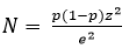
Where, N: size of the base sample; Z: 1.96, level of confidence (1- α)=95%; e: margin of error=5%; P: Prevalence (50% of prevalence was used for this purpose).
Fishes were captured live with nets or after emptying the pond. Fish collected as earlier described by Ngueguim DF et al, consisted of Clarias gariepinus (692), Cyprinus carpio (593) and Oreochromis niloticus (969). Fish samples were transported during the early hours (9:00-10:00) of the day in a sanitized flask with water from ponds Source to the Ichthyology and Applied Hydrobiology Laboratory of the University of Dschang, Cameroon [14].
Macroscopic examination
External anatomical examination: A clinical examination was carried out on the live or freshly dead fish. This involved the determination of any clinical signs of disease or abnormalities on the specimens as defined by Noga EJ et al [15]. External clinical signs (body color, scales, presence or absence of hemorrhage, lesions, etc) were observed on the field during a preliminary examination of live fish which were identified and recorded on a data sheet with a code for each fish. Additionally, each abnormality was photographed using a Sony digital camera at 10 mega pixels resolution. This was done in other to avoid inflammatory processes that make diagnosis uncertain or impossible.
Internal anatomical examination: At the laboratory, fishes were dissected by opening each specimen from the anus to the head with a sharp pair of scissors, taking care not to perforate the various organs as intestines. Then, the internal organs (liver and gonads) were removed with forceps and placed in petri dishes. Organs showing clinical sign of disease were placed in a container with 10% formalin for fixation and further histological examination.
Histological examination: The histological technique was carried out on the gonads and liver through the optimized protocol at the laboratory of Mountain University at Bangangté-Cameroon [5].
Parasitological examination of fish: In other to link abnormalities to fish parasites, parasitological examination was done. Hence, Standard parasitological procedures were used for fish examination and identification of ectoparasites and internal helminths respectively [14,16]. An infected fish sample was coded as 1 and uninfected as 0. Epidemiological parameters such as the prevalence (Pr) or infection rate and mean intensities (I) of infections were calculated according to Bush AO et al [16].
Statistical analysis: The data collected were subjected to different statistical analysis with the computer software Excel 2007, and SPSSv20. The chi-square test ( χ2) as well as the the obtained data was entered into Microsoft office Excel 2007 for descriptive statistics and transferred to the Statistical Package for the Social Sciences (version 22, SPSS Inc., USA) for further statistical analysis. The ecto and endoparasite prevalence rate calculated as the proportion of infected fish of the total number of fish examined and expressed as a percentage. The chi-square test was used to test significant levels within factors on prevalence rates and odds-ratios were determined for associated risk factors. Pearson correlation test were used to assess the relationship between the different variables and abnormalities observed. The significance level (p) considered was 5%.
Results
Typology and prevalence of external clinical signs of disease according to fish species
The typology and prevalence of external clinical signs of disease according to fish species are highlighted respectively in Figures 2 and 3. Irrespective of the fish species, a total of 7 abnormalities depending on their anatomical location were observed in 19.5% of the examined fishes. The prevalence of the clinical signs was higher in Cyprinus carpio (25.2%) followed by Oreochromis niloticus (17.5%) and Clarias gariepinus (17.3%). However, no significant difference was observed. Hemorrhage and erosion were about 9 times more frequent (p<0.05) than other abnormalities which accounted only for less than 2% each. Hemorrhage, erosion, absence of fin, deformation were common for fishes while red and protruding anus where specific to Cyprinus carpio and Oreochromis niloticus. Unlike Exolphtalmia and color alteration that were specific to Oreochromis niloticus and Cyprinus carpio respectively.

Figure 2: Typology of abnormalities: Note: (a) Erosion of the head in O. niloticus, (b) Erosion of tail fin in Clarias gariepinus; (c) Erosion of tail fin in C. carpio; (d) Body in C. gariepinus eroded; (e) Deformation of the head and body in C. gariepinus; (f) Deformation of the mouth of C. carpio.

Figure 3: Prevalence of some abnormalities recorded on farmed fish specimens in the West Region of Cameroon. Note: 
 Deformation;
  Red and protruding anus.
Prevalence of pathologies according to sex of cultured fishes
The prevalence of abnormalities according to the sex of fish as summarized in Table 1. It revealed that female fishes (20.1%) were the most affected compared to males (18.7%). However, no significant effect on the prevalence of abnormalities was recorded (p>0.05).
| Factors | Modalities | N (n) | Prevalence (%) | p-value (X2) |
|---|---|---|---|---|
| Sex | Males | 998(187) | 18.7 | 0.667 (0.185) |
| Females | 1256(252) | 20.1 |
Note: N: Number of fish examined; n: Number of fish with abnormalities.
Table 1: Prevalence of abnormalities according to fish sex
Typology and prevalence of internal clinical signs of disease related to fish species and organs
Gonad abnormalities: Overall, 2% of collected fishes showed Gonad abnormalities. Cyprinus carpio testis was asymmetrically developed and constricted while an ovary of Clarias gariepinus was found to be inflamed in the form of an oedema (Figure 4).

Figure 4: Gonad abnormalities observed in farmed fish from the West Region of Cameroon. Note: (A) Testicles with asymmetric development (black dash) and constriction of the gonad (red arrows) in Cyprinus carpio; (B) Inflammation of the female gonad in Clarias gariepinus.
Abnormalities in the liver: Liver alterations were recorded in 1.3% of Clarias gariepinus collected from fish farms in Menoua division (Figure 5).

Figure 5: Liver alterations in African catfish. Note: (A) Hepatic steatosis (fat black arrow); (B) Nodule inclusion (black arrows) in the liver; (C) Hepatic discoloration (Discoloured areas black arrows).
Histopathology of fish liver and gonads: The histological sections carried out on the fish livers revealed various tissue damages namely hemorrhagic necrosis while that of ovaries showed hermaphroditism and degeneration of the zona radiate (Figures 6, 7A and 7B).

Figure 6: Necrosis and haemorrhage in the fish liver 

Figure 7: Histopathology of the ovaries. Note: (A) Hermaphroditism in C. gariepinus:  (B) Degeneration of the zona radiata in O. niloticus.
Prevalence of ecto and endoparasites: A total of 894 (39.66%) fish were parasitized on the one hand by ectoparasites (34.87%) and on the other hand by endoparasites (8.47%) (Figure 8). 786 fish (34.87%; were infested with ectoparasites consisting of monogeneans (356; 15.79%), protozoa (356; 15.79%) and crustaceans (271; 12.02%). Monogenes consisted of Gyrodactylus sp. (285; 12.64%) and Dactylogyrus sp. (117; 5.19%). Protozoa consisted of Myxobolus sp. (232; 10.29%), Trichodina sp. (159; 7.05%) and Chidonella sp. (12; 0.53%). The crustaceans were composed of Branchiures comprising Argulus sp. (150; 6.65%) and Copepoda (150; 6.65%) including Lernea sp. (95; 4.21%) and Ergasilus sp. (55; 2.44%). Gyrodactylus sp. had the highest parasite infestation rate regardless of fish species while Chilodonella sp. had the lowest parasite infestation rate (Figure 8). As for the endoparasites, a total of 191 fish (8.47%) were parasitized by endoparasites including acanthocephalans (109; 4.84%) and nematodes (92; 4.08%). Acanthocephalus sp. (109; 4.84%) was the only acanthocephalan identified, while the Nematodes consisted of Camallanus sp. (27; 1.20%), Eustrongylides sp. (24; 1.06%), Capillaria sp. (43; 1.91%) and Orientatractis sp. (8; 0.35% [0.18-0.70]). Among these parasites, Acanthocephalus sp. achieved the highest parasite infestation rate for any fish species (Figure 8).

Figure 8: Prevalence of parasites identified in cultured fish in the West Cameroon Region.
Correlation between the prevalence of abnormalities and parasitism: The correlations between the prevalence of abnormalities and parasitism are shown in Table 2. Though all identified parasites except Chilodonella sp., Lernea sp., Ergasilus sp., Argulus sp., Eustrongylides sp and Orientatractis sp. were found on fish with abnormalities, no parasite was significantly (p>0.05) associated with the clinical signs of diseases observed on the host fish.
| Parasites | Exolphthalmia (N1 =5) |
Hemorrhage (N2 =428) | Erosion (N3 =406) |
Absence of fin (N4 =36) | Deformation (N5 =25) |
Red and protruding anus (N6 =14) |
Color alteration (N7 =14) | Abnormalities (N=440) |
||||||||
|---|---|---|---|---|---|---|---|---|---|---|---|---|---|---|---|---|
| n (%) | p | n (%) | P | n (%) | p | n (%) | p | n (%) | p | n (%) | p | n (%) | P | n (%) | p | |
| Monogens | ||||||||||||||||
| Gyrodactylus sp. | 0(0.0) | 1.000 | 6(1.3) | 0.476 | 6(1.4) | 0.857 | 1(0.3) | 0.207 | 0(0.0) | 0.648 | 0(0.0) | 1.000 | 0(0.0) | 1.000 | 6(1.3) | 0.472 |
| Dactylogyrus sp. | 0(0.0) | 1.000 | 21(0.5) | 0.332 | 2(0.5) | 0.448 | 1(0.2) | 0.361 | 0(0.0) | 1.000 | 0(0.0) | 1.000 | 0(0.0) | 1.000 | 2(0.5) | 0.328 |
| Protozoa | ||||||||||||||||
Myxobolus sp. |
0(0.0) | 1.000 | 6(1.3) | 0.491 | 7(1.8) | 0.571 | 1(0.3) | 0.201 | 0(0.0) | 0.651 | 1(0.2) | 0.295 | 0(0.0) | 1.000 | 6(1.3) | 0.474 |
| Trichodina sp. | 0(0.0) | 1.000 | 4(1.0) | 0.805 | 4(1.0) | 0.802 | 1(0.2) | 0.371 | 0(0.0) | 1.000 | 0(0.0) | 1.000 | 0(0.0) | 1.000 | 4(1.0) | 0.807 |
| Chilodonella sp. | 0(0.0) | 1.000 | 0(0.0) | 1.000 | 0(0.0) | 1.000 | 0(0.0) | 1.000 | 0(0.0) | 1.000 | 0(0.0) | 1.000 | 0(0.0) | 1.000 | 0(0.0) | 1.000 |
| Crustaceans | ||||||||||||||||
| Copepods | ||||||||||||||||
Lernea sp. |
0(0.0) | 1.000 | 0(0.0) | 0.625 | 0(0.0) | 0.636 | 0(0.0) | 1.000 | 0(0.0) | 1.000 | 0(0.0) | 1.000 | 0(0.0) | 1.000 | 0(0.0) | 0.620 |
| Ergasilus sp. | 0(0.0) | 1.000 | 0(0.0) | 0.625 | 0(0.0) | 0.636 | 0(0.0) | 1.000 | 0(0.0) | 1.000 | 0(0.0) | 1.000 | 0(0.0) | 1.000 | 0(0.0) | 0.620 |
| Branchiura | ||||||||||||||||
| Argulus sp. | 0(0.0) | 1.000 | 0(0.0) | 0.595 | 0(0.0) | 0.601 | 0(0.0) | 1.000 | 0(0.0) | 1.000 | 0(0.0) | 1.000 | 0(0.0) | 1.000 | 0(0.0) | 0.592 |
| Total Ectoparasites | 0(0.0) | 1.000 | 150(3.5) | 0.187 | 16(4) | 0.807 | 1(0.6) | 0.253 | 0(0.0) | 0.142 | 1(0.2) | 1.000 | 0(0.0) | 0.578 | 15(3.5) | 0.151 |
| Acanthocephala | ||||||||||||||||
| Acanthocephalus sp. | 0(0.0) | 0.979 | 257(0.6) | 0.283 | 2(0.5) | 0.713 | 0(0.0) | 1.000 | 0(0.0) | 1.000 | 0(0.0) | 1.000 | 0(0.0) | 1.000 | 3(0.6) | 0.480 |
| Nematods | ||||||||||||||||
| Camallanus sp. | 0(0.0) | 1.000 | 86(0.2) | 1.000 | 1(0.2) | 1.000 | 0(0.0) | 1.000 | 0(0.0) | 1.000 | 0(0.0) | 1.000 | 0(0.0) | 1.000 | 1(0.2) | 1.000 |
| Eustrongylides sp. | 0(0.0) | 1.000 | 0(0.0) | 0.222 | 1(0.2) | 0.699 | 0(0.0) | 1.000 | 0(0.0) | 1.000 | 0(0.0) | 1.000 | 0(0.0) | 1.000 | 0(0.0) | 0.223 |
| capillary sp. | 0(0.0) | 1.000 | 5(1.1) | 0.160 | 4(1.0) | 0.258 | 1(0.2) | 0.304 | 1(0.2) | 0.224 | 0(0.0) | 1.000 | 0(0.0) | 1.000 | 5(1.1) | 0.166 |
| Orientatractis sp. | 0(0.0) | 1.000 | 0(0.0) | 0.595 | 0(0.0) | 0.601 | 0(0.0) | 1.000 | 0(0.0) | 1.000 | 0(0.0) | 1.000 | 0(0.0) | 1.000 | 0(0.0) | 0.592 |
| Total Endoparasites | 0(0.0) | 1.000 | 8(1.9) | 0.341 | 7(1.8) | 0.399 | 1(0.2) | 1.000 | 1(0.2) | 1.000 | 0(0.0) | 1.000 | 0(0.0) | 1.000 | 8(1.9) | 0.452 |
| Total | 0(0.0) | 0.703 | 24(5.5) | 0.451 | 23(5.6) | 0.732 | 1(0.8) | 0.172 | 1(0.2) | 0.455 | 1(0.2) | 1.000 | 0(0.0) | 0.324 | 24(5.5) | 0.740 |
Note: N: Number of fish examined; n: Number of fish with abnormalities; p: probability of error.
Table 2: Correlation between fish abnormalities and parasitism
Correlation between endogenous factors and the occurrence of abnormalities observed in fish: The parasite prevalence was non-significantly (p>0.05) correlated with the appearance of the observed abnomalities (Table 3).
| Risk factors | r | p |
|---|---|---|
| Fish species | 0.002 | 0.483 |
| Sex | 0.017 | 0.334 |
| Parasite prevalence | -0.018 | 0.330 |
Note: r=correlation coefficients; p: probability of error.
Table 3: Correlation between endogenous factors and the occurrence of abnormalities observed in fish collected from farms in West Cameroon
Discussion
The results obtained with regard to anomalies corroborate those of Samia ASZ et al [7,9,17]. Indeed, exophthalmia has already been reported in fish by several authors. According to, Achat Soraya et Zarouri Samia et al, it was observed in the common carp Cyprinus carpio caught in the river at Tala Hamza [7]. Exophthalmia can be of infectious origin or linked to the supersaturation of the medium with gas [18]. Other causes of various origins have also been put forward, namely metabolic disorder, low oxygen content, after exposure of fish to ethanol and nutritional insufficiency (Vitamin A, E deficiency) [19-22]. Hemorrhage in fish is one of the abnormalities that reflect the degradation of the aquatic environment. Its presence is consistent with the descriptions reported by Girard P et al [19]. These authors related this type of abnormality to infectious agents, such as Enterobacteriaceae or viruses and protozoa, trauma, irritation or deficiency in Vitamin C, K [18,19,22,23]. It is also possible that excessive competition between fish will lead to a severe confrontation inducing various hemorrhages.
Deformation of fish is often associated with stunted growth and reduced swimming ability. Generally, deformation in fish is a multifactorial phenomenon and the determination of its origin is not obvious, but heavy metals can cause metabolic alterations that can act on bone metabolism and modify their mineralization [18,24]. But, the time of exposure and the degree of bioaccumulation of contaminants can increase the number of deformed fish [25]. According to Bogé G et al, contamination of the environment by heavy metals can be the cause of a deformation of the vertebral column in fish on the other hand linked this expression to an unsuitable diet and to vitamin deficiencies [26,27]. Moreover, hereditary phenomena are supposed to be at the origin [28]. However, points out that thermal deficits or chemical pollution can cause deformations of this type [29].
Fin erosion has already been reported by different authors for which various causes have been assumed such as: fungi and ectoparasites (Argulose argulus spp); cadmium; vitamin C deficiency, Vibrio anguillarum and predation [19,22,30,31]. Body erosion has also been linked to different sources of disturbance citing infestation by different agents such as bacteria and parasites, nutritional or vitamin deficiencies, unfavorable environmental factors and chemical pollution [19]. They later cause the degradation of the fish’s body mucus, which causes the disappearance of this protective layer. The affected organs are subsequently invaded by parasites or fungi, but also by viruses such as carp herpes virus 3 [18]. This is because the skin of fish represents an effective gateway for certain viruses [32]. These organs can also be affected by bacteria like Aeromonas sorbia detected in Garra rufa, since fish that are in a poor environment due to insufficient water quality such as high levels of nitrites, low levels of dissolved oxygen or high levels of carbon dioxide are more vulnerable to Aeromonas infection spp. [33]. Erosions and deformations can be linked to different physical factors such as: gas oversaturation, hypoxia, too low temperature, salinity, radioactivity, electric shocks, organochlorine compounds (pesticides, herbicides), heavy metals (Cd, Pb), parasites, bacteria, viruses or trauma (capture, predation). It can also be related to nutritional deficiencies, especially in vitamins [19], such as vitamins (A, C and D) [34]. Schàperclaus PW et al, showed that the exposure of fish to fertilizers (potash and ammonium salts) causes the separation of the fins [30].
Leclercq E et al, associates this change in phenotype (color alteration) during an established life stage, with the response to environmental interactions, but also with the transition between two stages of development phenotypically pre-adaptive to their ancestral ecosystems [20]. Color alterations in fish can be linked to several factors. Indeed, it can be of physiological origins, namely stress related to capture, but also a physical origin including, among others, hypoxia, excess CO2, gas oversaturation, insufficient mineralization of the water. It can also be of accidental origin (haemorrhage, trauma, irritation, blindness), genetic (hereditary diseases) or nutritional (vitamin deficiencies). Finally, a change in color can also be due to bacterial, parasitic (Myxosporidia, microsporidia) and viral infections [30,19].
It appears from this study that the anomalies identified would have a negative impact on the overweight of its host, probably by disturbing its physiology. The anomalies identified, although not significantly associated with the identified parasites, can be detrimental to fish production. Indeed, these superficial and/or deep lesions constitute entry points for secondary infections [35]. They can create serious dysfunctions for the individual which can lead to a drop in fertility [36]. Indeed, the clinical signs of naturally infested fish revealed respiratory distress, swimming at the surface and slow movements. These signs can be attributed to massive mucous secretions in the gills which can be used to dilute the irritation and act as a defense mechanism against infestation [37]. These results are almost similar to those found by who detected these abnormalities associated with the clinical signs mentioned above [36,38,39]. It would therefore be essential to monitor their appearance in fish farms in order to avoid such damage in fish farming.
Histopathological changes in the skin and gills of infested fish were recorded and the results are similar to those recorded by Aly HA et al [40]. Indeed, skin and gill lesions can be induced by feeding activity, attachment and locomotion of monogeneans causing massive destruction of respiratory epithelial cells and/or skin cells as well as ciliated trichodins on the skin. These results agree with the work of Gado MS et al [41,42]. In the gills, hyperplasia takes place between the gill lamellae and the parasites then feed on the newly produced cells and damage the gill tissues. Thus, the branchial epithelium is completely destroyed, leaving large denuded areas between the filaments. Perforated basement membrane and capillaries cause bleeding. This condition disrupts breathing [42]. Eventually, the fish die of suffocation [42]. The physical presence of Trichodina sp. and Myxobolus sp localized in the vessels damages the gills and opens the gateway for secondary infection. This leads to high mortality in the nursery operating system in carp [43]. The gills were frayed, congested and slimy with a mottled appearance. The mottled appearance can be attributed to the destruction of efferent vessels. This leads to rapid occlusions of the vessel and then a thrombus forms resulting in ischemia, which in turn leads to necrosis [36,41]. Other cases showed pale gills with destruction of gill filaments. These results are consistent with those reported by Eissa AE et al [44,45]. Additionally, Vinoth R et al, found that infested fish had extremely pale gills, indicating that the gills were seriously affected [46]. Histopathological findings from the affected intestines showed massive destruction, necrosis, atrophy as well as leukocyte infiltration. These results are in agreement with the work of [45,47]. These results can be attributed to the parasite-induced mechanical injury and its feeding on the delicate tissues on the circulating blood resulting in hemorrhage. Slow, rapid blood clot, thrombus, ischemia, finally necrosis and widespread damage to the gastrointestinal tract [48-50]. In view of the impact of parasites at the tissue level, identifying fish diseases and their causative agents is necessary to initiate preventive measures against pathogens. It would therefore be essential to monitor the appearance of parasites in fish farms given the growing involvement of fish farming on a daily basis.
Conclusion
Several anomalies have been recorded on the external and internal anatomy of the fish. However, they were not significantly associated with parasites. The histological sections performed showed various tissue and cellular alterations in the affected organs. It will be advisable to extend these studies to other agroecological zones of Cameroon with a view to establishing a map of the biosecurity measures, prevalence of recurrent pathologies in fish farms according to the seasons, and the means of prevention and control of these pathologies. This will contribute effectively to the increase of national fish farming production. It will equally be useful to carry out additional work to that of the present study, notably a microbiological study on the different organs of the affected fish, and a microbiological study of the fish water, as well as a nutritional analysis of the rations distributed to farmed fish.
Acknowledgement
The authors are grateful to the Staff of MINEPIA and fish farmers of the West region of Cameroon for their generous cooperation.
Conflict of Interest
There is no conflict of interest.
References
- Kaleem O, Sabi AF. Overview of aquaculture systems in Egypt and Nigeria, prospects, potentials, and constraints. Aquac Fish. 2021;6(6):535-547.
- Adah AD, Lawal S, Oniye JS, Ola-Fadunsin SD, Okunbanjo OO, Adah AS. Occurrence and risk factors associated with Eimeria species infections in Clarias gariepinus and Heteroclarias species. 2022;Niger. J. Parasitol
- Muringai RT, Mafongoya P, Lottering RT. Sub-Saharan Africa freshwater fisheries under climate change: A review of impacts, adaptation, and mitigation measures. Fishes. 2022;7(3):131.
- Alves LM, Correia JP, Lemos MF, Novais SC, Cabral H. Assessment of trends in the Portuguese elasmobranch commercial landings over three decades (1986–2017). Fish Res. 2020;230:105648.
- Djoudad-Kadji H, Benslimane S, Chevalier C, Kadji B, Exbrayat Jm, Iguer-Ouada M. Visualisation des coupes histologiques des follicules ovariens de barbus callensis variation de fixateurs et de colorants. Rev. fr. histotechnol. 2011;24(1):21-28.
- Marcon L, Bazzoli N, Honor Mounteer A, Anjos Benjamin LD. Histological and histometric evaluation of the liver in Astyanax bimaculatus (Teleostei: Characidae), exposed to different concentrations of an organochlorine insecticide. Anat Rec Hoboken. 2015;298(10):1754-1764. [Crossref]
[Google Scholar] [PubMed]
- Samia ASZ. Aspects anatomo-histopathologiques de quelques espèces de poissons d’eau douce dans la région de Bejaia. Mémoire de Fin de Cycle en vue de l’obtention du diplôme de Master en Environnement et Santé publique. Université A. MIRA-Bejaia. Faculté des Sciences de la Nature et de la Vie, Algérie. 2017.84.
- Kadji H. Caractérisation de la reproduction du poisson d'eau douce Barbus barbus callensis au niveau de l'Oued Soummam dans la région de Béjaïa.
- Gasmi A, Ouari K. Aspects anatomo-histopathologique de quelque espèce de poissons d’eau douce dans la région de Béjaia. Travail de diplôme en vue de l’obtention du diplôme master, Filière science biologique université A. MIRA.Béjaia, 2017.45.
- Arezzouk A. Evaluation des paramètres biologiques et environnementaux liés au cycle de vie de quelques espèces de poissons d’eau douce vivant dans le barrage TICHI-HAF (Béjaia). Travail de diplôme en vue de l’obtention du diplôme master, Filière science biologique université A. MIRA. Béjaia.2017.43.
- Uhland FC, Hélie P, Higgins R. Infections of Edwardsiella tarda among brook trout in Quebec. J. Aquat Anim Health. 2000;12(1):74-77.
[Crossref] [Google Scholar] [PubMed]
- IRAD. "Rapport annuel des activités." Institut de Recherche Agricole pour le Développement, Yaounde, Cameroon. 2013.
- Thrusfield M. Veterinary epidemiology. John Wiley & Sons. 2018.
- Ngueguim DF, Kouam MK, Tiogue CT, Miegoue E, Feumba AK, Zebaze LB, et al. prevalence and associated risk factors of ectoparasite infections of cultured fish species in the west region of Cameroon. Int j fish aquat sci. 2020;8(3):310-320.
- Noga EJ. Fish disease: Diagnosis and treatment. John Wiley & Sons. 2010.
- Bush AO, Lafferty KD, Lotz JM, Shostak AW. Parasitology meets ecology on its own terms: Margolis et al. revisited. J Parasitol.1997;83(4):575-583.
[Crossref] [Google Scholar] [PubMed]
- Touahria sonia. Etude anatomohistopathologique de quelques espèces de poissons d’eau douce (BEJAIA). Mémoire de Fin de Cycle en vue de l’obtention du diplôme de Master de Biochimie appliquée. Université A. MIRA-Bejaia, Faculté des Sciences de la Nature et de la Vie, Algérie.2018:78.
- Richard Y, Baillargeon JP, Masse H. Guide de classification des anomalies externes des poissons d’eau douce du Québec. ENSEMBLE. 2016:186.
- Girard P, Elie P. Manuel d’indentification des principales lésions anatomomorphologiques et des principaux parasites externes des anguilles. Collection étude Cemagref bordeaux n° 110. 2007: 81.
- Leclercq E, Taylor JF, Migaud H. Morphological skin colour changes in teleosts. Fish Fish. 2010;11(2):159-193.
- Ali S, Champagne DL, Alia A, Richardson MK. Large-scale analysis of acute ethanol exposure in zebrafish development: A critical time window and resilience. PloS one. 2011;6(5):e20037.
- Tacon AG. Pathologie nutritionnelle des poissons. Signes morphologiques des carences et intoxications alimentaires chez les poissons d'elevage. Food and Agriculture Org. 1995.
- Vigier JF. Les pathologies des anguilles: snythese des connaissances sur la pathologie chez les differentes especes du genre Anguilla. 1997:200.
- Louiz I, Menif D, Ben Attia M, Ben Hassine OK. Incidence des déformations squelettiques chez trois espèces de Gobiidae de la lagune de Bizerte (Tunisie). Cybium. 2007;31(2):209-216.
- Kim SK, Lee DS, Oh JR. Characteristics of trophic transfer of polychlorinated biphenyls in marine organisms in Incheon North Harbor, Korea. Environ Toxicol Chem. 2002;21(4):834-841.
[Crossref] [Google Scholar] [PubMed]
- Bogé G, Roche H, Houvet D. Les indicateurs physiologiques de toxicité en milieu marin. Océanis. 1991;17(4):351-365.
- Labat R, Roqueplo C, Ricard JM, Lim P, Burgat M. Actions écotoxicologiques de certains métaux (Cu-Zn-Pb-Cd) chez les poissons dulçaquicoles de la rivière Lot. InAnnales de Limnologie. Int J Limnol. 1977; 13(2):191-207.
- Brusle J. Skeletal abnormalities in fish and their multifactorial aetiology: A review. In E.A.F.R, seventh international conference. Diseases of fish and shellfish. 1995:90.
- Mellinger DK. Optimized detection of sperm whale clicks. Proc. 2nd Intl. Workshop on Detection and Localization of Marine Mammals Using Passive Acoustics. Monaco. 2005:37.
- Schàperclaus PW. Les maladies des poissons « maladies non parasitaires » Bulletin français de pisciculture trente-cinquième année. N°206.1962 :5-17.
- Breuil G. Vibriosis in sea bass. ICES Identification Leaflets for Diseases and Parasites of Fish and Shellfish/Fiches d'identification des maladies et parasites des poissons, crustacés et mollusques. 1991;50:1-4.
- Michel B, Fournier G, Lieffrig F, Costes B, Vanderplasschen A. Cyprinid herpesvirus 3. Emerg Infect Dis. 2010;16(12): 1835-1843.
[Crossref] [Google Scholar] [PubMed]
- Majtán J, Černy J, Ofúkaná A, Takáč P, Kozánek M. Mortality of therapeutic fish Garra rufa caused by Aeromonas sobria. Asian Pac J Trop Biomed. 2012;2(2):85-87.
[Crossref] [Google Scholar] [PubMed]
- Hamdouni Y, Dhaouadi R. Journée Nationale sur la valorisation des résultats de la Recherche dans le domaine de la Pêche et de l’Aquaculture, Tunisie, Suivi sanitaire et étude histologique des gonades du tilapia du Nil, Oreochromis niloticus, dans un élevage en circuit fermé.2014:87-90.
- Fomena A. Les Myxosporidies et Microsporidies des poissons d’eau douce du Sud–Cameroun: Étude faunistique, Ultrastructure et Biologie (Doctoral dissertation, Thèse de Doctorat d’État. Université de Yaoundé I:397.
- Eissa IA. Parasitic fish diseases in Egypt. Dar El-Nahda El-Arabia Publishing. 2002;32:149-160.
- Yambot AV, Lopez EA. Gill parasite, Lamproglena monodi Capart, infecting the Nile tilapia, Oreochromis niloticus L., cultured in the Phillipines. InProceedings of the 3rd Symposium on Diseases in Asian Aquaculture. Manila: Asian Fisheries Society. 1997:175-177.
- Ragias V, Tontis D, Athanassopoulou F. Incidence of an intense Caligus minimus Otto 1821, C. pageti Russel, 1925, C. mugilis Brian, 1935 and C. apodus Brian, 1924 infection in lagoon cultured sea bass (Dicentrarchus labrax L.) in Greece. Aquaculture. 2004;242(1-4):727-733.
- Woo PT. Fish diseases and disorders: Volume 1: Protozoan and metazoan infections. USA: CAB International, London.2006: 466-565
- Aly HA, Abdel-Rahim MM, Lotfy AM, Abdelaty BS, Sallam GR. The applicability of activated carbon, natural zeolites, and probiotics (EM®) and its effects on ammonia removal efficiency and fry performance of European seabass Dicentrarchus labrax. J Aquac Res. 2016;7(11):459-466.
- Noor El-Deen AI. Comparative studies on the prevailing parasitic diseases in monosex tilapia and natural male tilapias in Kafr El Sheikh Governorate fish farms (Doctoral dissertation, Ph. D. Thesis, Fac. Vet. Med., Kafr El-Sheikh University). 2017.
- Gado MS, Mahfouz NB, Moustafa EM, Lolo EE. Prevalence of some ectoparasitic diseases in African catfish (Clarias gariepinus) at Kafr El-Sheikh governorate. International Journal of Fsheries and Aquatic Studies. 2017;5(3):576-583.
- Subashinghe R. Disease’s control and health management in aquaculture. FAO Aquaculture Newsletter 1995; 9: 8-11.
- Eissa AE, Hussein HA, Zaki MM. Detection of avian influenza (H5N1) In some fish and shellfish from different aquatic habitats across some Egyptian provinces. Life Sci. 2012;9(3):2702-2712.
- Mohamed AE, Hassan MA, Osman HA. Protochondracanthus alatus infesting gills of some marine fish species. Global Veterinaria. 2013;11(4):406-413.
- Vinoth R, Ajith Kumar TT, Ravichandran S, Gopi M, Rameshkumar G. Infestation of Copepod parasites in the food fishes of Vellar estuary, Southeast coast of India. Acta Parasitologica Globalis. 2010;1(1):1-5.
- Khidr AE, Samak OA, Said AE, Ghoneim AM, Fahmy SA. Structural and functional observations on the appendages of gill parasite, Lernanthropus Kroyeri (Copepoda: Lernanthropidae) Infesting the sea bass Dicentrarchus Labrax. Nature and Science. 2014;12(2):101-107.
- Eissa AE, Moustafa M, Abdelaziz M, Ezzeldeen NA. Yersinia ruckeri infection in cultured Nile tilapia, Oreochromis niloticus, at a semi-intensive fish farm in lower Egypt. Afr J Aquat Sci. 2008;33(3):283-286.
- Purivirojkul W, Areechon N. A survey of parasitic copepods in marine fishes from the Gulf of Thailand, Chon Buri Province. Agric Nat Resour. 2008;42(5):40-48.
- Mohammed SY. Histopathological changes in the intestines and gonad of grouper fish Epinephelus microdon infected with nematode parasites, Red Sea Coast, Sudan. 2017.
Citation: Ngueguim DF, Kouam MK, Fonkwa G, Katte HB, Nack J, Awah-Ndukum J (2023) Endogenous Factors Associated to Anatomo-Pathological Lesions of Cultured Fish Species in the West Region of Cameroon. J Aquac Res Dev. 14:807.
Copyright: © 2023 Ngueguim DF, et al. This is an open-access article distributed under the terms of the Creative Commons Attribution License, which permits unrestricted use, distribution, and reproduction in any medium, provided the original author and source are credited.