Indexed In
- The Global Impact Factor (GIF)
- CiteFactor
- Electronic Journals Library
- RefSeek
- Hamdard University
- EBSCO A-Z
- Virtual Library of Biology (vifabio)
- International committee of medical journals editors (ICMJE)
- Google Scholar
Useful Links
Share This Page
Journal Flyer

Open Access Journals
- Agri and Aquaculture
- Biochemistry
- Bioinformatics & Systems Biology
- Business & Management
- Chemistry
- Clinical Sciences
- Engineering
- Food & Nutrition
- General Science
- Genetics & Molecular Biology
- Immunology & Microbiology
- Medical Sciences
- Neuroscience & Psychology
- Nursing & Health Care
- Pharmaceutical Sciences
Research Article - (2021) Volume 20, Issue 9
Comparison of the Synergistic Effect of Vacuum Therapy or Ultrasound Associated with Low Power Laser Applied in Temporomandibular Disorders
Vitor Hugo Panhóca1*, Patricia Eriko Tamae1,2, Antonio Eduardo Aquino Jr1 and Vanderlei Salvador Bagnato12Central Paulista University Centre-UNICEP/São Carlos, São Paulo, Brazil
Abstract
This work aims to investigate and compare the effects of new technologies that can help in the rehabilitation process of people with Temporomandibular Disorders (TMD) by controlling pain and reducing the inflammatory process, increasing joint functionality and improving their quality of life patients. Twenty female and male volunteers, aged between 18 and 55 years, with joint or muscle TMD were selected. The volunteers were randomly divided into 2 groups: Group USL-Treated with Ultrasound (US) plus Laser and Group VL-treated with Vacuum plus Laser (VL). For the clinical study, 2 therapeutic sessions were carried out during 4 weeks. Clinical evaluations were performed: anamnesis and diagnosis; pain assessment using an analogue pain scale; assessment of range of motion (total opening of the mouth) and assessment of quality of life (OHIP-14). For statistical analysis, the Kolmogorov-Smirnov normality test was performed, followed by a one-way ANOVA analysis, using the Student-Newman-Keuls test for parametric data. The results consist of reduced pain, increased range of motion of the Temporomandibular Joint (TMJ) and improved quality of life in both groups. We can conclude that laser combined with US or vacuum are effective in the treatment of TMD.
Keywords
Low-level laser therapy, Vacuum therapy, Ultrasound, Temporomandibular disorder, OHIP-14.
Introduction
Temporomandibular Dysfunction (TMD) has been a clinical situation very present in dental clinics and offices. Often, the prescription medication and use of mandibular stabilizing plates are sufficient to soften acute conditions, but do not have a rapid effect for certain conditions that become chronic. TMD is characterized by functional and pathological changes that affect TMJ, masticatory muscles and, eventually, other parts of the stomatognathic system. Approximately 40% of the Brazilian population has some sign or symptom of TMD, and about 5% of the total requires treatment and control of this disease [1]. TMD affects adults and children, which can lead to loss of quality of life and labor damage [2]. The adult female population between 20 and 50 has the highest prevalence of TMD, with a proportion of 5:1 compared to men [1].
The etiology of TMD is multifactorial. Traumas of the mandible or temporomandibular joint (TMJ), anxiety, nocturnal bruxism, emotional stress, changes in masticatory muscles due to micro trauma or caused by continuous parafunctional habits, rheumatic conditions and postural abnormalities may be related to the development of TMD [3]. Complementary alternative therapies we can see the application of Ultrasound (US) and vacuum therapy for the treatment of muscle and joint dysfunctions. Therefore, laserbased systems as a light source to promote analgesia and tissue deinflammation can be combined with the application of US and vacuum therapy with anti-inflammatory synergistic effect and pain relief in TMD and mastication muscles to obtain greater speed and therapeutic efficiency.
Therefore, noninvasive therapies that dispense with the use of systemic medication appear as an interesting, non-aggressive option with minimal risk of side effects [4]. The use of Laser and Led has been demonstrated in previous research by our research group (Biophotonics Laboratory-CEPOF-IFSC-USP)as complementary therapy in the treatment of TMD [5,6] and other morbidities, such as: cervical dentinal hypersensitivity, trigeminal neuralgias, post-surgical paresthesia and facial paralysis [7]. In addition to our studies, other previous studies published are found in the international literature applying light source with low power, red or infrared laser with the objective of treatment of TMDs and obtained positive results in this treatment [8].
In view of these positive results and protocols already pre-established for TMD laser treatment, this work shows the synergistic use of Ultrasound (US) and vacuum therapy in the complementary treatment of TMD in order to optimize these treatments. In this context, in a broader view, the rehabilitation process with the use of physical, mechanical and electromagnetic agents; would aim to control pain and improve joint function, muscles and tendons with reduction of inflammatory process and stiffness, as well as increased range of motion, muscle strength, autonomy and improved quality of life.
Material and Methods
Twenty volunteers were selected and randomly evaluated in the dental office at the Biophotonics Laboratory of the Institute of Physics of São Carlos, University of São Paulo (IFSCUSP). For TMD treatment, volunteers were selected according to the "Research Diagnostic Criteria for Temporomandibular Disorders (RDC/TMD)" designed by Dworkin [9].
This study was approved by the ethics committee for human studies of the Irmandade da Santa Casa de Misericórdia de São Carlos, C.A.A.E.09096219.0.0000.8148. All doubts expressed by the participants were clarified orally, and it was explained that the participation in the study was voluntary. All volunteers who participated in the experiment read and signed the informed consent form (TCLE). This study is registered in the Brazilian Clinical Trials Registry (ReBEC) under the clinical trial registration number RBR-4g6dc35 with the identifiers:
• CAAE issuing body: Brazil Platform 09096219.0.0000.8148
• WHO International Clinical Trials Registry platform: UTN code U1111-1266-6024.
• Approval number of the clinical research ethics committee (from Santa Casa de Misericórdia de São Carlos): 3.244.307.
Clinical measurements
Oral opening and pain assessment measures using a Visual Analog Scale (VAS) from 0 to 3, 0=pain-free, 1=comfort with mild pain, 2=acute pain only during stimulus application and 3=acute pain during stimulus application and continuous after removal; were evaluated in three moments: pre-treatment (T0), after the eight treatment sessions (T1) and 30 days after the end of treatment (T2).
The Oral Health Impact Profile (OHIP-14), validated for Portuguese, was used for quality-of-life analysis in all patients [10]. This instrument consists of 14 questions that assess 7 domains: functional limitation, physical pain, psychological distress, physical limitation, psychological limitation, social limitation and disability. The score is determined using a 5-point scale: 0=never, 1=rarely, 2=sometimes, 3=often,and 4=always. The maximum score of all answers to the 14 questions is equivalent to 56 points; the higher the score, the lower the quality of life. The OHIP instrument was applied in three moments: T0=pre-treatment and T1=post-treatment.
Groups and devices used in this study
Patients in the USL group were treated with the Recupero® (MM Optics, São Carlos, SP–Brazil) device, which consists of applying US and laser to the same system (Figure 1A). The wavelengths applied were laser diode at 808 nm, power of 100 mW and spot area (output) 1.76 mm2; and US with frequency of 1.0 MHz, intensity 1W/cm2, 50% of pulsed duty cycle and effective radiation area of 1.6 cm2 (Table 1). Transparent water-based gel was used as a means of transmission for the active us tip. The synergistic treatment with the US and laser were applied simultaneously for 120 seconds per region to the masseter muscle, anterior temporal muscle and TMT on the left and right sides of the face, with slow and smooth circular movements.
| Parameters | |
|---|---|
| Laser diode | 808 nm |
| Power | 100 mW |
| Irradiance | 5.7 W/cm2 |
| Energy density/area | 684 J/cm2 |
| Energy density/session | 4.104 J/cm2 |
| Total energy density | 32.832 J/cm2 |
| Energy/area | 12D |
| Spot size | 1.76 mm2 |
| US (Recupero®) | |
| Frequency | 1.0 MHz |
| Effective radiation | 1.6 cm2 |
| Intensity (%) | 1 W/cm2 |
| Mode pulsed | 50% |
| VL (VacumLaser®) | |
| Negative pressure | 100-150 mbar |
| Mode pulsed | MP7=40 pulses/min. |
| Energy/area/laser | ≤72 J*/ region |
Table 1: Photobiomodulation therapy and ultrasound or vacuum parameters used in synergistic treatment.
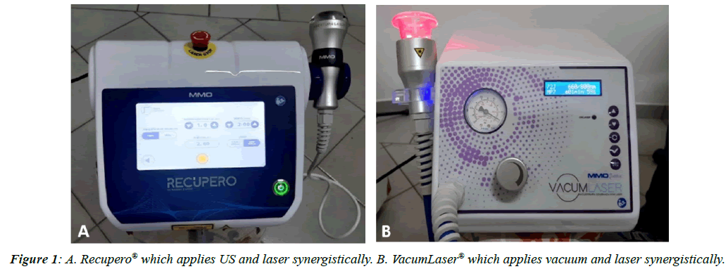
Figure 1: A. Recupero® which applies US and laser synergistically. B. VacumLaser® which applies vacuum and laser synergistically.
The VL group was treated with the VacumLaser® device (MM Optics, São Carlos, SP–Brazil) that synergistically applies low-power laser and negative pressure through suction cup in a system shown in Figure 1B. All volunteers received the same treatment. The treatments applied were with lowpower laser with 6 laser diode outputs being 3 to 808 nm and 3 to 660 nm wavelength concomitantly; each laser output has the power of 100 mW producing a total power of 600 mW; and laser spot area is 1.76 mm2. The suction cup was applied punctually to the face synergistically to the laser, as shown in Figures 2 and 3 on both sides of the face. In case of unilateral pain, the application was also unilateral. The suction cups were adjusted with negative pressure from 100 to 150 mbar and in pulsed mode MP7=40 pulses per minute. The suction cup used was the small one with a diameter of 40 mm. During all application of VacumLaser® suction cups vegetable oil was previously applied to the patient's facial surface so that the suction cup could slide over the skin tissue. The application was made for 120 seconds per region.
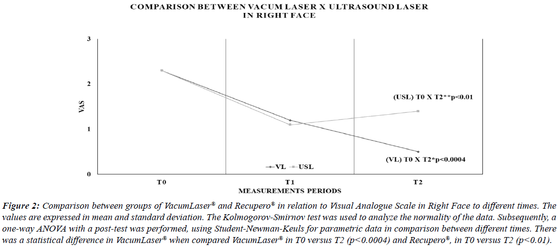
Figure 2: Comparison between groups of VacumLaser® and Recupero® in relation to Visual Analogue Scale in Right Face to different times. The values are expressed in mean and standard deviation. The Kolmogorov-Smirnov test was used to analyze the normality of the data. Subsequently, a one-way ANOVA with a post-test was performed, using Student-Newman-Keuls for parametric data in comparison between different times. There was a statistical difference in VacumLaser® when compared VacumLaser® in T0 versus T2 (p<0.0004) and Recupero®, in T0 versus T2 (p<0.01). Figure 3:
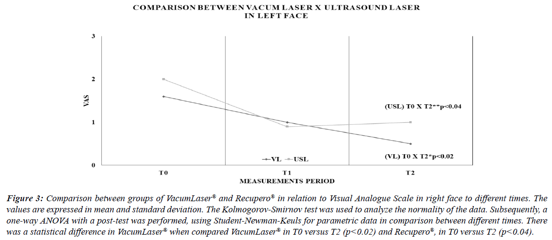
Figure 3: Comparison between groups of VacumLaser® and Recupero® in relation to Visual Analogue Scale in right face to different times. The values are expressed in mean and standard deviation. The Kolmogorov-Smirnov test was used to analyze the normality of the data. Subsequently, a one-way ANOVA with a post-test was performed, using Student-Newman-Keuls for parametric data in comparison between different times. There was a statistical difference in VacumLaser® when compared VacumLaser® in T0 versus T2 (p<0.02) and Recupero®, in T0 versus T2 (p<0.04).
All patients participated in two sessions per week for 4 weeks, a total of eight sessions. The frequency and number of sessions were stipulated based on the protocol used in previous studies by our research group [5,6].
Results
The results of pain evaluation were fragmented in relation to the right and left facial hemispheres and can be seen in Figures 2 and 3 respectively and at 3 different times, being T0=initial values, T1=values after treatment and T2=values 30 days after treatment. When we observed Figure 2, we found the comparison of the analog pain scale compared in the 3 different times, in relation to the variables of vacuum laser and US laser, analyzing the hemisphere of the right face. In this comparison, it is possible to observe the significant difference between the initial values (T0) versus the values of 30 days after treatment (T2) for vacuum laser (p<0.0004) and ultrasound laser (p<0.01) (Figure 2).
When observing Figure 3, the comparison of the analog pain scale of the left facial hemisphere shows, at the 3 different times, in relation to the variables of vacuum laser and ultrasound laser, the significant difference between the initial values (T0) versus the values of 30 days after treatment (T2) for vacuum laser (p<0.02) and ultrasound laser (p<0.04) (Figure 3).
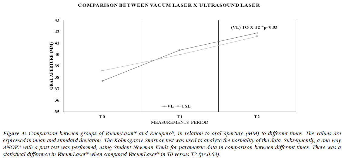
Figure 4 shows the comparison between the maximum oral opening, when the variables vacuum laser and ultrasound laser were observed, in the 3 different times. It is possible to analyze the best maximum buccal opening, when the vacuum laser variable is used in the comparison between the initial times (T0) versus after 30 days (T2), for p<0.03. When we observed the use of the ultrasound laser variable, there was no significant difference (Figure 4).
Figure 4: Comparison between groups of VacumLaser® and Recupero®, in relation to oral aperture (MM) to different times. The values are expressed in mean and standard deviation. The Kolmogorov-Smirnov test was used to analyze the normality of the data. Subsequently, a one-way ANOVA with a post-test was performed, using Student-Newman-Keuls for parametric data in comparison between different times. There was a statistical difference in VacumLaser® when compared VacumLaser® in T0 versus T2 (p<0.03).
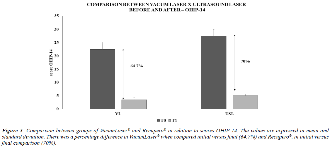
The results evaluated using the OHIP-14 questionnaire show that both groups had an improvement in quality of life, and the VL group had an improvement of 64.7%, while the USL group achieved 70% improvement (Figure 5).
Figure 5: Comparison between groups of VacumLaser® and Recupero® in relation to scores OHIP-14. The values are expressed in mean and standard deviation. There was a percentage difference in VacumLaser® when compared initial versus final (64.7%) and Recupero®, in initial versus final comparison (70%).
Discussion
TMD, of myofascial origin, consists of episodes of pain with periods of exacerbation and remission, and may be persistent in some cases. It is commonly associated with the presence of trigger points and discomfort occurs as a response to tension and painful alteration of the muscle and fascia, which can be local or referred to with sensitivity and pressure on palpation [11]. The main disorder in patients with TMD is related to motor problems caused by changes in muscle activity and not to joint problems. Numerous therapeutic interventions are potentially effective in the treatment of TMD such as mandibular stabilizer plates, low-power laser, therapeutic ultrasound, Transcutaneous Electrical Stimulation (TENS), as well as manual therapy techniques and exercises [11].
In this context, the use of associative technologies, which provide the conjugated use of therapies, previously unique, are increasingly being used for pain in its various spectrums, such as TMD, osteoarthritis [12], fibromyalgia [13] and Parkinson's [14]. Thus, therapeutic ultrasound combined with therapeutic laser and vacuum therapy, conjugated to therapeutic laser, stand out. It is established in the literature that therapeutic ultrasound has properties that enable the transmission of energy and mechanical absorption generating effects the increase of vascularization [6], enzymatic activity and collagen synthesis, as well as accelerating the inflammatory process for tissue repair, for example, the skin, muscle, cartilage and bone. In addition, there is an increase in the speed of neural conduction and the nociceptive threshold that contribute to the treatment of pain as well as promotes the change of muscle contractility [15-17], with consequent reduction of spasms, generating osteogenic stimulation through the potentials generated by deformation (SGPs) and acceleration of tissue repair of muscle lesions [17,18].
The therapeutic laser has photobiomudulation with pain effect, healing, anti-inflammatory [19] as well as analgesic effect [20]. In addition, it causes increased vascularization,formation of fibroblasts, among other biomolecules [19].
Vacuum therapy, is a therapy that consists of sucking the skin through negative pressure through suction cups with the intention of increasing local blood flow and can be applied for stimulating effect of lymphatic drainage, muscle relaxation after therapy by activating circulation by suction, improving the circulatory system, promoting anti-inflammatory effect and relief of muscle, joint and tendon pain [21].
Thus, the present study aimed to compare these conjugated technologies as a focus on pain reduction and mouth opening amplitude. Thus, it was observed in Figures 2 and 3 the decrease in pain in both facial hemispheres and in both conjugated technologies. However, a better statistical result was observed when the conjugated vacuum therapy technology associated with therapeutic laser was used, pointing out that the anti-inflammatory and analgesic action, when associated with mechanical, stimulator and vascular action, allows a greater reduction of pain, when compared to the action of therapeutic ultrasound and therapeutic laser conjugated therapies.
Regarding the amplitude of mouth opening (Figure 4), it was observed that the action of vacuum therapy and therapeutic laser (p<0.03) was also better than the therapeutic ultrasound conjugate. In this comparison, the mechanical, stimulator and vascular effects associated with analgesic and anti-inflammatory actions were fundamental, promoting greater relaxation of the area and, in this case, stimulating a greater mouth opening.
When observing Figure 5, where the values of OHIP-14 are shown, indicating the improvement in quality of life, it is possible to record the improvement in quality of life in both therapies compared. Although the action of therapeutic ultrasound and therapeutic laser was briefly better (70%), in relation to the action of vacuum therapy and therapeutic laser, (64%), its proximity is positive, showing that both have high results in improving quality of life.
The results presented allow a clear observation of the positive action of conjugated therapies, with greater emphasis on the conjugated action of vacuum therapy and therapeutic laser, allowing greater pain reduction, greater amplitude of mouth opening and, consequently, increasing the quality of patients' lives. The synergy of therapies is established as an efficient means for the treatment of TMD, performed in a few sessions, in a noninvasive or drug-free manner.
Conclusions
Conjugated therapies are increasingly promising in the treatment of pain and inflammation. Conjugated therapy with the use of negative pressure and therapeutic laser proved to be efficiently in the treatment of TMD, allowing pain reduction, improvement of mouth opening amplitude and consequent improvement in quality of life. Noninvasive and non-pharmacological therapeutic actions are increasingly present and the treatment of TMD is present in this context efficiently and soon intervention.
Evaluating the results obtained in this study, we can say that laser therapy combined synergistically with vacuum or ultrasound improved the quality of life of patients with TMD as seen in the evaluation through the OHIP-14 questionnaire. The laser combined with vacuum or ultrasound can be considered as complementary or alternatives treatment to control pain and increase oral opening in patients with TMD.
Acknowledgments
The authors acknowledge the financial support by Financier of Studies and Projects (FINEP)–grant 01.13.0430-00, São Paulo Research Foundation (FAPESP)-grant no. 2013/14001- 9 (CEPOF–CEPID Program) and has the partnership with MM Optics.
REFERENCES
- de Godoi Gonçalves DA, Dal Fabbro AL, Campos JA, Bigal ME, Speciali JG. Symptoms of temporomandibular disorders in the population: an epidemiological study. J Orofac Pain. 2010;24(3).
- Graue AM, Jokstad A, Assmus J, Skeie MS. Prevalence among adolescents in Bergen, Western Norway, of temporomandibular disorders according to the DC/TMD criteria and examination protocol. Acta Odontol Scand. 2016;74(6):449-455.
- Karthik R, Hafila MF, Saravanan C, Vivek N, Priyadarsini P, Ashwath B. Assessing prevalence of temporomandibular disorders among university students: a questionnaire study. J Int Soc Prev Community Dent. 2017;7(Suppl 1):S24.
- Butts R, Dunning J, Pavkovich R, Mettille J, Mourad F. Conservative management of temporomandibular dysfunction: A literature review with implications for clinical practice guidelines (Narrative review part 2). J Bodyw Mov Ther. 2017;21(3):541-548.
- Panhoca VH, Lizarelli RD, Nunez SC, de Andrade Pizzo RC, Grecco C, Paolillo FR, et al. Comparative clinical study of light analgesic effect on temporomandibular disorder (TMD) using red and infrared led therapy. Lasers Med Sci. 2015;30(2):815-822.
- Panhóca VH, Lopes LB, Paolillo FR, Bagnato VS. Treatment of temporomandibular disorder using synergistic laser and ultrasound application. Oral Health Dent Manag. 2018;17:1-5.
- Panhóca VH, Nogueira MS, Bagnato VS. Treatment of facial nerve palsies with laser and endermotherapy: a report of two cases. Laser Phys Lett. 2020;18(1):015601.
- Xu GZ, Jia J, Jin L, Li JH, Wang ZY, Cao DY. Low-level laser therapy for temporomandibular disorders: a systematic review with meta-analysis. J Pain Res. 2018;2018.
- LeResche L, Von Korff MR. Research diagnostic criteria for temporomandibular disorders: review, criteria, examinations and specifications, critique. J Craniomandib Disord. 1992;6(4):301-355.
- Santos CM, Celeste RK, Hilgert JB, Hugo FN. Testing the applicability of a model of oral health-related quality of life. Cad Saude Publica. 2015;31:1871-1880.
- Schmitter M, Rammelsberg P, Hassel A. The prevalence of signs and symptoms of temporomandibular disorders in very old subjects. J Oral Rehabil. 2005;32(7):467-473.
- de Souza Simão ML, Fernandes AC, Ferreira KR, de Oliveira LS, Mário EG. Comparison between the singular action and the synergistic action of therapeutic resources in the treatment of knee osteoarthrosis in women: A blind and randomized study. J Nov Physiother. 2019;9(411):2.
- Junior AE, Carbinatto FM, Franco DM, Bruno JS, Simão ML. The Laser and Ultrasound: The Ultra Laser like Efficient Treatment to Fibromyalgia by Palms of Hands–Comparative Study. J Nov Physiother. 2020;11(447):2.
- Tamae PE, Santos AV, Simão ML, Canelada AC, Zampieri KR. Can the associated use of negative pressure and laser therapy be a new and efficient treatment for parkinson's pain? A comparative study. J Alzheimers Dis Parkinsonism. 2020;10(488):2.
- Lee W, Kim H, Lee S, Yoo SS, Chung YA. Creation of various skin sensations using pulsed focused ultrasound: evidence for functional neuromodulation. Int J Imaging Syst Technol. 2014;24(2):167-174.
- Srbely JZ, Dickey JP. Randomized controlled study of the antinociceptive effect of ultrasound on trigger point sensitivity: novel applications in myofascial therapy? Clin Rehabil. 2007;21(5):411-417.
- Chen KC, Lin AC, Chong CF, Wang TL. An overview of point-of-care ultrasound for soft tissue and musculoskeletal applications in the emergency department. J Intensive Care. 2016;4(1):1-1.
- Manbachi A, Cobbold RS. Development and application of piezoelectric materials for ultrasound generation and detection. Ultrasound. 2011;19(4):187-196.
- Chen CH, Hung HS, Hsu SH. Low‐energy laser irradiation increases endothelial cell proliferation, migration, and eNOS gene expression possibly via PI3K signal pathway. Lasers in Surgery and Medicine: J Am Lasers Surg Med. 2008;40(1):46-54.
- Karu T. Photobiological fundamentals of low-power laser therapy. IEEE J Quantum Electron. 1987;23(10):1703-1717.
- Volpi A. Analysis of the efficacy of vacuum therapy in the treatment of fibro geloid edema through thermography and biophotogrammetry. Physioter Bras. 2017;11:70–77.

