Indexed In
- The Global Impact Factor (GIF)
- CiteFactor
- Electronic Journals Library
- RefSeek
- Hamdard University
- EBSCO A-Z
- Virtual Library of Biology (vifabio)
- International committee of medical journals editors (ICMJE)
- Google Scholar
Useful Links
Share This Page
Journal Flyer

Open Access Journals
- Agri and Aquaculture
- Biochemistry
- Bioinformatics & Systems Biology
- Business & Management
- Chemistry
- Clinical Sciences
- Engineering
- Food & Nutrition
- General Science
- Genetics & Molecular Biology
- Immunology & Microbiology
- Medical Sciences
- Neuroscience & Psychology
- Nursing & Health Care
- Pharmaceutical Sciences
Research Article - (2024) Volume 23, Issue 3
Association between cephalometric maxillary incisorâs inclination and facial and growth axes.
Samar Bou Assi1*, Ziad Salameh2, Antoine Hanna1, Roula Tarabay1 and Anthony Macari12Department of Dental Medicine, Lebanese University, Beirut, Lebanon
Received: 13-Jan-2020, Manuscript No. OHDM-24-3110; Editor assigned: 16-Jan-2020, Pre QC No. OHDM-24-3110 (PQ); Reviewed: 30-Jan-2020, QC No. OHDM-24-3110; Revised: 02-Sep-2024, Manuscript No. OHDM-24-3110 (R); Published: 30-Sep-2024, DOI: 10.35248/2247-2452.24.23.1114
Abstract
With the increased awareness to facial esthetics in general and to smile esthetics more specifically, most individuals seeking orthodontic treatment are looking for perfection in both the look of their teeth and the attractiveness of their smile. In this regard, the evaluation of the inclination of maxillary anterior teeth during orthodontic treatment constitutes a routine practice to ascertain an adequate positioning in the basal bone and in relation to the facial features. More specifically, the assessment of the Maxillary Incisors’ inclination (MI) and position are major aspects of orthodontic treatment planning, judging treatment progress and determining treatment outcome.
KeywordsFacial esthetics, Orthodontic treatment, Maxillary incisors, Inclination.
Introduction
The axial inclination of the maxillary incisors can be determined by several means and methods and can be assessed according to different reference lines and planes. The diagnostic records that provide information on incisors’ position are the conventional lateral cephalograph and the articulated dental casts. Whether evaluated on lateral cephalometric radiographs and/or dental casts, the orthodontist must ensure the appropriate position of the maxillary incisors by evaluating their axial inclination at the beginning of treatment, by assessing it periodically throughout treatment and by attaining a final position that would be judged as most esthetic and appropriate for the patient [1]. Traditionally, cephalometric assessment of the inclination of the maxillary incisors is performed by measuring the angles between the long axis of the incisor (joining incisal tip to apex) and planes 4-13: Sella-Nasion (SN), the Frankfort Horizontal plane (FH), the Palatal Plane (PP), APogonion (A-Pog), N-Pogonion (N-Pog), N-A line and the line parallel to N-perpendicular passing through point A, to the maxillary occlusal plane, to the bony orbit, to the forehead and glabella as well as the inter-incisal angle. These measurements are used, taking into consideration all the variables that might affect them [2].
While the cephalometric inclination of the maxillary incisors has been extensively studied, little has been said about its potential association with facial pattern, namely facial axes. In Ricketts’ analysis, the Facial Axis (FA) angle (mean 90° ± 3.5°) is the angle formed between Ba-N plane and the line extending from foramen rotundum (Pt) to constructed gn. A smaller angle suggests a retruded position of the chin, whereas an angle greater than 90° suggests a protrusive or forward growing chin. In other words, if the facial axis is greater than normal, the mandibular growth is in a forward direction and if it is smaller than normal, the vector of growth is in a more downward than forward direction. According to Ricketts, facial axis to Na-Ba angle does not change with growth. Similarly, the Growth Axis (GA) f (described by Downs, as the angle between Sella turcica (S) to Gnathion (Gn) line and Frankfort Horizontal line), ranges from a minimum of 53° to a maximum of 66°, with a mean reading of 59.4° ± 3.8°. This angle indicates the growth pattern of the mandible [3].
In our routine cephalometric assessment, we have been observing that well inclined maxillary incisors are often close to being parallel to the facial axis. Thus the objectives of this study were to:
• Determine if there is an association between the inclination of the maxillary incisors and facial and growth axes in an orthodontic population.
• Assess the degree of parallelism between the maxillary incisors’ long axis and facial and growth axes.
• Study the possibility of introducing a new cephalometric assessment of the maxillary incisors by using the facial and/or growth axes as an individual reference.
Materials and Methods
500 consecutive lateral cephalograms, were selected from patients’ data at the department of orthodontics and dentofacial orthopedics at the American university of Beirut. Available lateral cephalometric x-rays of growing and adult patients taken prior to or at the end of orthodontic treatment (after removal of brackets) placed according to the natural head position at an appropriate distance (sagittal plane-film distance of 13 cm) were studied. We excluded patients with craniofacial [4].
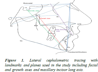
The 500 lateral cephalograms were digitized using the dolphin orthodontic software (dolphin imaging and management solutions, La Jolla, CA). Angular measurements were computed to determine the inclination of maxillary I to SN, PP and NA, as well as FA, GA and maxillary I to NBa and true horizontal. Measurements of different variables were done on the digitized lateral cephalograms (Figure 1) [5].
Parallelism between maxillary incisors and facial axis was then evaluated by measuring the angle between Nasion-Basion line (N/Ba) and the long axis of maxillary incisors and the angle between FA and N Ba. The FA and maxillary incisors were also measured relative to the true horizontal [6].
Parallelism between maxillary incisors and growth axis evaluated by measuring the angle between N Ba and long axis of maxillary incisors and the angle between GA and N Ba. The GA and maxillary incisors were also measured relative to the true horizontal (Figure 1).

Figure 1: Lateral cephalometric tracing with landmarks and planes used in the study including facial and growth axes and maxillary incisor long axis.
Statistical analyses
Data cleaning was performed on all entered data to check for potential errors done during data entry. An initial frequency distribution was generated for all variables to check for any potential outliers. Data was stratified based on the inclination of maxillary incisors and descriptive statistics were computed between four cephalometric measurements assessing the position of the well positioned maxillary I (Maxillary I to Facial Axis (FA), maxillary I to SN, maxillary I to NA and maxillary I to Palatal Plane (PP) [7].
Associations were tested using chi-square tests for categorical data and Pearson‘s correlation along with the ANOVA test for independent samples followed by the Bonferroni post-hoc test for continuous data. For all parameters, two-sided p-values were reported. P-value <0.05 was considered as statistically significant. All analyses were completed using IBM SPSS Statistics version 24.
Results
Inter-rater reliability was calculated on all variables of randomly chosen cephalograms (n=50) that were digitized by a second examiner. Intra-class correlation coefficients were high (>0.9) (Table 1) [8].
| Groups (n=498) | A (n=143) | B (n=214) | C (n=141) | ANOVA (p) | Comparisons among groups | |||||
|---|---|---|---|---|---|---|---|---|---|---|
| Mean | SD | Mean | SD | Mean | SD | A-B | A-C | B-C | ||
| Age (y) | 22.92 | 12.675 | 17.01 | 9.214 | 17.5 | 9.527 | <0.001 | <0.001 | <0.001 | NS |
| Cephalometric measurements | ||||||||||
| Facial axis/NBa | 87.513 | 5.6443 | 88.164 | 4.969 | 89.592 | 6.274 | 0.006 | NS | 0.005 | NS (0.055) |
| Facial axis/horiz | 115.692 | 12.7348 | 116.886 | 4.1211 | 118.011 | 3.8681 | 0.038 | NS | 0.032 | NS |
| Y axis/NBa | 92.622 | 5.9075 | 92.196 | 7.9898 | 94.301 | 5.2341 | 0.013 | NS | NS | 0.012 |
| Y axis/horiz | 122.188 | 3.4613 | 121.535 | 3.446 | 122.426 | 3.504 | 0.042 | NS | NS | NS (0.055) |
| U1-NBa | 71.469 | 6.4144 | 84.773 | 4.2958 | 94.84 | 5.5137 | <0.001 | <0.001 | <0.001 | <0.001 |
| U1-horiz | 101.332 | 6.3739 | 113.561 | 3.9916 | 123.011 | 4.7971 | <0.001 | <0.001 | <0.001 | <0.001 |
| I/NA | 9.19 | 5.6815 | 22.433 | 3.0814 | 31.986 | 3.9108 | <0.001 | <0.001 | <0.001 | <0.001 |
| I/PP | 98.694 | 5.731 | 110.864 | 10.4596 | 120.833 | 4.8464 | <0.001 | <0.001 | <0.001 | <0.001 |
| I/SN | 89.126 | 6.6481 | 102.717 | 7.0901 | 112.922 | 5.3128 | <0.001 | <0.001 | <0.001 | <0.001 |
Table 1. Means of age, selected cephalometric measurements in groups stratified on maxillary incisors inclination.
The angle facial axis/NBa was statistically significantly different (p=0.005) between groups A (retroclined maxillary incisors) and C (proclined maxillary incisors), being more increased in group C (89.6° ± 6.2°). Similarly, facial axis/ horizontal was statistically difference between group A and C, more increased in group C than the other two groups (118° ± 3.9°). In addition, the Y axis to NBa and to the horizontal were statistically significantly different between the three groups with p=0.013 and 0.042 respectively (Table 1). Maxillary incisors to NBa and to the horizontal were also statistically significantly different between the groups A, B and C (p<0.001).
Statistically significant positive correlations between facial and growth axes on one hand and maxillary incisors inclination on the other hand, existed in the total sample (Table 2). While groups A and B did not correlate with facial and growth axes, interestingly the group C of proclined maxillary incisors had moderate positive correlation to facial and growth axes angles to NBa and to the horizontal. In group C, maxillary incisors to SN correlated the highest to facial axis/horizontal (r=0.401; p<0.001) and to both facial axis/NBa (r=0.31; p<0.001) and to growth axis/NBa (r=0.34; p<0.001) [9].
| Groups | U1/NA | U1/PP | U1/SN | U1/NaBa | U1/H | ||||||
|---|---|---|---|---|---|---|---|---|---|---|---|
| r | p-value | r | p-value | r | p-value | r | p-value | r | p-value | ||
| Facial axis/NaBa | A | 0.121 | NS | 0.12 | NS | 0.222 | 0.008 | 0.349 | <0.001 | -0.097 | NS |
| B | -0.057 | NS | 0.077 | NS | 0.094 | NS | 0.457 | <0.001 | -0.179 | 0.009 | |
| C | 0.172 | 0.042 | 0.104 | NS | 0.313 | <0.001 | 0.488 | <0.001 | 0.07 | NS | |
| ALL | 0.158 | <0.001 | 0.157 | <0.001 | 0.219 | <0.001 | 0.333 | <0.001 | 0.081 | NS | |
| Facial axis/Horizontal | A | 0.019 | NS | 0.017 | NS | 0.027 | NS | 0.052 | NS | 0.077 | NS |
| B | -0.077 | NS | 0.016 | NS | 0.048 | NS | 0.115 | NS | 0.123 | NS | |
| C | 0.327 | <0.001 | 0.233 | 0.005 | 0.401 | <0.001 | 0.327 | <0.001 | 0.328 | <0.001 | |
| ALL | 0.121 | 0.007 | 0.104 | 0.02 | 0.135 | 0.003 | 0.149 | 0.001 | 0.157 | <0.001 | |
| Growth axis/NaBa | A | 0.057 | NS | 0.066 | NS | 0.077 | NS | 0.285 | 0.001 | -0.151 | NS |
| B | -0.054 | NS | 0.055 | NS | 0.072 | NS | 0.271 | <0.001 | -0.149 | 0.03 | |
| C | 0.147 | NS | 0.12 | NS | 0.339 | <0.001 | 0.541 | <0.001 | 0.038 | NS | |
| ALL | 0.087 | NS | 0.107 | 0.013 | 0.139 | 0.002 | 0.236 | <0.001 | 0.021 | NS | |
| Growth axis/Horizontal | A | 0.137 | NS | 0.073 | NS | 0.097 | NS | 0.18 | 0.03 | 0.092 | NS |
| B | -0.08 | NS | 0.032 | NS | 0.096 | NS | 0.224 | 0.001 | 0.151 | 0.027 | |
| C | 0.178 | 0.035 | 0.158 | NS | 0.274 | 0.001 | 0.242 | 0.004 | 0.302 | <0.001 | |
| ALL | 0.045 | NS | 0.054 | NS | 0.092 | 0.041 | 0.121 | 0.007 | 0.1 | 0.025 | |
Table 2. Correlations between maxillary incisors’ inclination and facial/growth axes.
Discussion
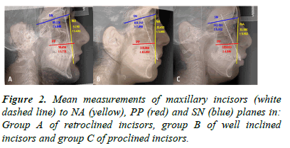
The main contribution of this study was the association between maxillary incisors and growth and facial axes inclinations. Patients with proclined maxillary incisors have increased facial and growth axes to cranial base angles (Figure 2). This finding suggests that when anterior rotation of the lower face (mainly the mandible) occurs, the maxillary incisors tend to procline further. In addition, this compensatory mechanism follows in the opposite direction with individuals exhibiting decreased facial and growth axes angles, whereby the maxillary incisors are more retroclined [10]. To the best of our knowledge, this was the first time such an association was evaluated.
Our sample was constituted of growing and non-growing subjects.

Figure 2: Mean measurements of maxillary incisors (white dashed line) to NA (yellow), PP (red) and SN (blue) planes in: Group A of retroclined incisors, group B of well inclined incisors and group C of proclined incisors.
Growth/compensatory issues
The growth axis indicates the growth pattern of the mandible. If the growth axis is within normal range, it means that the mandible is growing down and forward (normodivergent pattern); if it is larger than the normal, the mandible has a vertical vector of growth (hyperdivergent pattern) and if smaller than normal, the vector of growth is in a more horizontal direction (hypodivergent pattern). A small facial axis suggests a retropositioned chin, whereas an angle greater than 90 degrees suggests a protrusive or forward growing chin [11]. In other words, if the facial axis is greater than normal, the mandibular growth is in a forward direction and if it is smaller than normal, the vector of growth is in a more downward than forward direction. The findings of this study are concomitant with that of cephalometric studies assessing the maxillary incisors’ compensation in different mandibular sagittal and vertical positions. However, our sample was classified on maxillary incisors’ inclination irrespective of sagittal or vertical relationship between the jaws. In other words, groups A, B and C of retroclined, well inclined and proclined maxillary incisors were randomly chosen. In this regard, the three groups include class I, II and III malocclusions, as well as hypo-, normo- or hyper-divergent mandibles.
Clinical implications
The findings of this study suggest that position of the maxillary incisors may be cephalometrically evaluated in reference to facial and growth axes. Furthermore, more compensation of the maxillary incisors to one end may be acceptable in a parallel way to facial axis inclination [12].
Research issues
We stratified maxillary inclination in three groups based on three angles of the incisors to the anterior cranial base, palatal plane and Nasion-A plane. Thus, one individual can be classified in one of the three groups only if all three angles read one diagnosis of either retroclined, well inclined or proclined. In this method, we were confirming the position of the maxillary incisor to be the same whether evaluated to the maxillary basal, cranial base or to the position of the maxilla in the profile. Rarely though we find that those three angles read one outcome, thus the importance of this comparison within this large sample of subjects.
It would be interesting to longitudinally follow patients while they are growing to evaluate changes in the association between maxillary incisors inclination and facial axis. Nevertheless and given that mandibular plane rotation to cranial base endures minimum changes throughout growth, our sample of non-treated growing and non-growing individual’s stands as a valid one to evaluate the above-mentioned association. Furthermore, no differences existed between the groups when stratified on gender.
While radiographic evaluation is a common approach for the assessment of maxillary incisors’ inclination, some clinicians evaluate this inclination on dental casts.
These practitioners consider that the use of the lateral cephalograph for that purpose is sometimes difficult and prone to errors caused by radiographs digitization. In our study, dental casts were discarded because of possible inappropriate trimming and because they’re not useful in identifying the degree of maxillary incisors’ proclination or retroclination [13].
Conclusion
Maxillary incisors tend to be more proclined in individuals with more increased facial and growth axes compared to subjects with average facial and growth axes inclinations. Moderate positive correlation existed between maxillary incisors and facial and growth axes. Higher correlations were found in the group of proclined incisors. The findings suggest that inclination of the maxillary incisors may be compensatory to the growth pattern with a more leeway of compensation to procline than to retrocline. Further longitudinal investigation on growing and non-growing subjects would be beneficiary to confirm this potential compensatory mechanism.
References
- Richmond S, Klufas ML, Sywanyk M. Assessing incisor inclination: A non-invasive technique. Eur J Orthod. 1998;20(6):721-726.
[Crossref] [Google Scholar] [PubMed]
- Ghahferokhi AE, Elias L, Jonsson S, Rolfe B, Richmond SRolfe B, Richmond S. Critical assessment of a device to measure incisor crown inclination. Am J Orthod Dentofacial Orthop. 2002;121(2):185-191.
- Knosel M, Kubein-Meesenburg D, Sadat-Khonsari R. The third-order angle and the maxillary incisor's inclination to the NA line. Angle Orthod. 2007;77(1):82-87.
[Crossref] [Google Scholar] [PubMed]
- Ellis III E, McNamara Jr JA. Cephalometric evaluation of incisor position. Angle Orthod. 1986;56(4):324-344.
- Houston WJ. The analysis of errors in orthodontic measurements. Am J Orthod. 1983;83(5):382-390.
[Crossref] [Google Scholar] [PubMed]
- Baumrind S, Frantz RC. The reliability of head film measurements: 1. Landmark identification. Am J Orthod. 1971;60(2):111-127.
[Crossref] [Google Scholar] [PubMed]
- Haraguchi S, Iguchi Y, Takada K. Asymmetry of the face in orthodontic patients. Angle Orthod. 2008;78(3):421-426.
[Crossref] [Google Scholar] [PubMed]
- Brodie AG. The growth of the jaws and the eruption of the teeth. Oral Surg Oral Med Oral Pathol. 1948;1(4):334-341.
[Crossref] [Google Scholar] [PubMed]
- Schlosser JB, Preston CB, Lampasso J. The effects of computer-aided anteroposterior maxillary incisor movement on ratings of facial attractiveness. Am J Orthod Dentofacial Orthop. 2005;127(1):17-24.
- Sassouni V. A roentgenographic cephalometric analysis of cephalo-facio-dental relationships. Am J Orthod. 1955;41(10):735-764.
- Assi SB, Macari A, Hanna A, Tarabay R, Salameh Z. Cephalometric evaluation of maxillary incisors inclination, facial and growth axes in different vertical and sagittal patterns: An original study. J Int Soc Prev Community Dent. 2020;10(3):292-299.
[Crossref] [Google Scholar] [PubMed]
- Bhattacharya A, Bhatia A, Patel D, Mehta N, Parekh H, Trivedi R. Evaluation of relationship between cranial base angle and maxillofacial morphology in Indian population: A cephalometric study. J Orthod Sci. 2014;3(3):74-80.
[Crossref] [Google Scholar] [PubMed]
- Assi SB, Macari A, Hanna A, Aybout J, Salameh Z. Comparison between pre and posttreatment inclination of maxillary incisors in adults: Association with facial and growth axes. Contemp Clin Dent. 2022;13(4):344-348.
[Crossref] [Google Scholar] [PubMed]
Citation: Assi SB, et al. Association between cephalometric maxillary incisor’s inclination and facial and growth axes. Oral Health Dent Manage. 2024;23(3):1114.
