Citations : 2345
Dentistry received 2345 citations as per Google Scholar report
Indexed In
- Genamics JournalSeek
- JournalTOCs
- CiteFactor
- Ulrich's Periodicals Directory
- RefSeek
- Hamdard University
- EBSCO A-Z
- Directory of Abstract Indexing for Journals
- OCLC- WorldCat
- Publons
- Geneva Foundation for Medical Education and Research
- Euro Pub
- Google Scholar
Useful Links
Share This Page
Journal Flyer

Open Access Journals
- Agri and Aquaculture
- Biochemistry
- Bioinformatics & Systems Biology
- Business & Management
- Chemistry
- Clinical Sciences
- Engineering
- Food & Nutrition
- General Science
- Genetics & Molecular Biology
- Immunology & Microbiology
- Medical Sciences
- Neuroscience & Psychology
- Nursing & Health Care
- Pharmaceutical Sciences
Research Article - (2024) Volume 14, Issue 3
Assessment of Efficacy of PAP, Acridine Orange and AGNOR in Detecting Micronuclei in Oral Exfoliative Smears in Smokers-A Cross Sectional Comparative Study
D Anupriya*, Harini Priya A. H, R Sathish Muthukumar and Sreeja CReceived: 06-Jul-2020, Manuscript No. DCR-24-5311; Editor assigned: 09-Jul-2020, Pre QC No. DCR-24-5311 (PQ); Reviewed: 23-Jul-2020, QC No. DCR-24-5311; Revised: 01-Aug-2024, Manuscript No. DCR-24-5311 (R); Published: 29-Aug-2024, DOI: 10.35248/2161-1122.24.14.696
Abstract
Cigarette smoking is the major cause of oral cancer, which accounts for around two-thirds of all reported cases of oral squamous cell carcinomas annually. Tobacco use in any form is considered as a behavioural process which elicits physiological and psychologic addictive mood among users. Smoking leads to various adverse health effects. A cigarette contains over 4000 identifiable chemical carcinogens, with 200 known carcinogens that show toxicity. Comparing other anatomic sites of our body, the oral cavity shows high tendency for developing malignancy. This is because, oral cavity acts as the first barrier in the route of inhalation or ingestion of food and it can metabolize procarcinogens contained in tobacco smoke to reactive products. This may be due to the nature of the oral mucosa which is made of stratified non keratinized epithelia, which allows the superficial cells to maintain an intact structure and in turn promoting colour absorption and easy observation of nuclear characteristics under the microscope.
Keywords
Smokers; Cigarette smoking; PAP; AgNOR
Introduction
Among the various cytological structures, micronucleus plays a more promising role in early detection of dysplastic changes. Recently, the concept of detecting micronuclei has become an upgraded topic in the study of oral cancer. The origin of micronuclei is from the chromosome fragments or whole chromosomes, which are formed during the metaphase/anaphase of mitosis (cell division). This micronuclei can be a whole lagging chromosome (aneugenic event leading to chromosome loss) or an accentric chromosome fragment detaching from a chromosome after breakage (clastogenic event) which does not combine with the daughter nuclei. The carcinogenic and mutagenic agents and the nitrosamines present in the cigarettes are believed to be responsible for the formation of micronuclei. Various studies were performed regarding micronuclei which showed the correlation of frequency of micronuclei and severity of the genotoxic damage and the severity of the condition can be measured in terms of grading the lesion. Analysis of micronuclei in the oral exfoliated cells can indicate the amount of malignant transformation because, any genetic damage like micronuclei formation seen in these basal cells is reflected in the exfoliated cells. The most commonly used stains for micronuclei are PAP, Feulgen stains, Giemsa stains. PAP has the capacity to stain various layers differentially. Thus, in this study, we compared the staining efficacy of acridine orange, PAP and AgNOR for micronuclei in exfoliated oral mucosal cells of smokers and fractal dimension of micronuclei from each stain is calculated in order to analyse the morphological changes undergone by the epithelial cells to transform into neoplastic cell.
Materials and Methods
This study aims to compare the efficacy of PAP, acridine orange and AGNOR in detecting micronuclei, to evaluate the frequency of micronuclei in smears of exfoliated cells obtained from two groups i.e., smokers and non-smokers and fractal dimension analysis was also done to analyze the morphological changes. Patient visiting the department of oral pathology and oral medicine will be recruited for the study. 30 patients with history of smoking cigarettes were selected as the study group and 30 healthy individuals with no history of habits were selected as the control group [1]. Case history will be recorded along with thorough oral examination. The inclusion criteria for the study group were followed pertaining to Fagerstrom index. Patients with Fagerstrom score of 5 and above was selected. Before collection of samples, written consent was obtained from the individuals. To remove the oral debris, the patients were asked to rinse their mouth with mouthwash thoroughly prior to sample collection. The buccal mucosa was scraped gently using a broad, moistened wooden spatula. The cells obtained were immediately smeared on pre-cleaned labeled slides which is then fixed by placing in Coplin jars containing 90% alcohol. Three slides were obtained from each subject of the study group. The 3 stains were prepared.
Acridine orange: 50 mg acridine orange is dissolved in 10 ml of distilled water to prepare a stock solution and stored in the refrigerator. 1 ml of acridine orange stock solution and 0.5 ml of glacial acetic acid is added to 50 ml of distilled water to prepare a working solution.
The slide is put in the coplin jar containing acridine orange staining working solution (0.01%). After 2 minutes of staining, the slides are washed gently with water and dried and then treated with xylene [2]. The slides were then examined in a fluorescent microscope.
AgNOR: Composition of silver colloid developer included solution A-(Silver nitrate: 50 gms, deionised water: 100 ml) and solution B-(Gelatin: 2 gms, formic acid: 1 ml, deionised water: 100 ml). Working solution is solution A: 2 part and solution B: 1 part.
The slides were stained with freshly prepared silver colloidal solution (1 part by volume of 2% gelatin in 1% formic acid and two parts by volume of 50% aqueous silver nitrate solution) in a closed coplin jar for 35 min at room temperature. A dark environment was maintained throughout the reaction time. Then the slides were washed with double distilled ionized water. Later treated with 5% sodium thiosulphate for 5 minutes and washed in double distilled deionized water and dried.
PAP: The slides were dipped in 1% acetic acid (10 dips). Then treated with Harris’s haematoxylin, preheated 60°C. Wash in tap water. Dip in 1% acetic acid. Then treat with OG-6. Followed by 1% acetic acid (10 dips) and in EA-50 [3]. Again dip in 1% acetic acid and methanol 10 dips xylene 10 dips.
The slides were then subsequently stained with PAP, acridine orange stains and AgNOR. From each slide, 100 exfoliated cells were viewed under 400 × magnification in light microscope (PAP and AgNOR) and in fluorescent microscope at 525 nm wavelength (acridine orange). The count of micronuclei in each of those cells was recorded and later the photographs of the micronuclei from each slide was collected and run through the image software to calculate the fractal dimension.
Results
Assessment of micronuclei
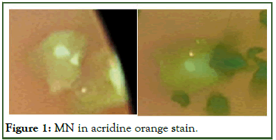
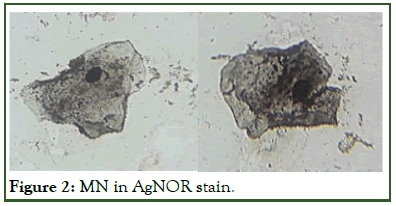
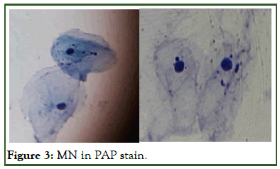
The micronuclei were counted in each slide and the average was calculated. The test results obtained are given in Tables 1 and 2 showing the mean values of 21.1, 15.6 and 26 in smokers group for acridine orange, AgNOR and PAP respectively. Additionally, the D value for fractal dimension was calculated which was 1.29, 1.16, 1.11 for acridine orange, AgNOR and PAP respectively [4]. Comparing the mean values, the value was the highest for PAP as compared to other stains. Hence, the study concludes that there is significant difference in the staining property of the micronuclei by the different stains in the smokers’ group (Figures 1-3).
| Group 1 | Group 2 |
|||
|---|---|---|---|---|
| Normal PAP | Smokers-Acridineorange | Smokers-AgNOR | Smokers-PAP | |
| Total no. of MN | 71 | 633 | 469 | 782 |
| Average | 2.3 | 21.1 | 15.6 | 26 |
Table 1: Camparison of no. of MN and mean of the three stains.
| D value of acridine orange | D value of AgNOR | D value of PAP |
|---|---|---|
| 1.29 | 1.16 | 1.11 |
Table 2: Fractal D values.

Figure 1: MN in acridine orange stain.

Figure 2: MN in AgNOR stain.

Figure 3: MN in PAP stain.
Discussion
The reactive products of cigarette are carcinogenic such as polycyclic hydrocarbons and nitrosamines which are metabolized and results in the production of epoxide and oxidative combustion [5]. The micronuclei are fragments of chromosomes or whole chromosomes that were lost during cell mitosis especially the anaphase stage because of a clastogenic eventwhich causes chromosomal breakage-or an aneugenic eventfragmentation of spindle fibres. The term micronucleus was first proposed in early 1970's by Schimdt, Boller and Heddle. They demonstrated it as a reliable indicator of genotoxic potential of mutagens after in vivo exposure using bone marrow erythrocytes in animal subjected studies. These micronuclei are formed due to the genotoxic and carcinogenic components present in tobacco, alcohol, betel nut, etc which are largely consumed by the general population. According to a study, a significant correlation was observed with increased frequencies of micronuclei and cigarette smoking [6]. So these micronuclei can be considered as indicators of genotoxic damage and chromosomal aberrations which can detect early stagecarcinogenesis.
Kashyap, et al., have proposed distinct advantages of the micronuclei assay over other cytological techniques which accounts for readily accessible site of sample collection from multiple areas of a particular site in the same patient, noninvasive technique and feasibility in conducting longitudinal epidemiological studies. Another study by Suhas, et al., showed the significant correlation between smoking and micronuclei.
Palaskar S and Jindal C compared PAP and Giemsa staining techniques to detect micronuclei in exfoliated buccal mucosal cells and concluded that PAP is a better stain over Giemsa for micronucleus detection.
In the present study conducted, micronuclei were observed and scored using three different staining procedures using PAP, acridine orange and AgNOR. PAP is a multichromatic stain which is used widely for oral exfoliated cells. It is mainly known for its superior background clarity in smears, free of debris and other proteinaceous material [7]. Acridine orange is a cellpermeable, nucleic acid selective dye that emits green fluorescence when bound to dsDNA (at 520) and red fluorescence when bound to ssDNA or RNA (at 650 nm). Since it is a cationic dye, it also enter acidic compartments such as lysosomes which in low pH conditions, will emit orange light. Nucleolar Organiser Regions (NORs) are defined as nucleolar components containing a set of argyrophilic proteins, which are selectively stained by silver methods. After silver-staining, the NORs can be easily identified as black dots exclusively localised throughout the nucleolar area and are called "AgNORs". The results of the study showed that smokers show an overall increase in frequency of micronuclei when compared to nonsmokers. PAP was found to give superior results in detecting micronuclei when compared with other stains. Acridine orange inspite of showing lower frequencies of micronuclei, was found to give fewer chances of false positive result [8]. This is because the stain binds and fluoresce only the nuclear component whereas the other DNA non specific stains may also stain the keratin granules, dye granules, bacterial contaminants which can lead to false positive result.
Fractal geometry is a tool used to characterize irregularly shaped and complex figures. One of the advantages of fractal analysis is the ability to quantify the irregularity and complexity of objects with a measurable value, which is called the fractal dimension [9]. It can be determined using the box-counting method. During the past years’ fractal analysis has been applied also in tumor pathology to characterize irregular boundaries of tumors and their nuclei. In this study, the images of each slide of all the 3 stains were taken was converted into binary images. The box length (e) and the number of boxes N (e) required to cover the structures of the nuclei was recorded [10]. If an object is an ideal fractal, N (e) increases and the fractal dimension can be determined as the slope of a double-logarithmic plot of N (e) over e.
It was noted that, even if the objects have the same fractal dimension, they might differ in their texture which can be evaluated by a parameter known as lacunarity. Fractal analysis has the potential for providing a measure of irregularity in shape of nuclei and can also reflect the corresponding functional status of the cell [11].
The results of the study conducted concluded that smokers show an overall increase in frequency of micronuclei when compared to non-smokers; also, PAP was found to give superior results in detecting micronuclei with relative accuracy in comparison with acridine orange and AgNOR stains [12]. But considering the minimal difference in the mean value, acridine orange also gives a better quality in expressing a micronuclei.
Conclusion
Micronuclei formation indicates genotoxic and cytotoxic changes in the oral mucosa which is a common dysplastic change seen in oral cancerous cells. The presence of micronuclei in exfoliated cells of smokers can be attributed as an early dysplastic marker for disease onset and progression. When comparing PAP, acridine orange and AgNOR it was found that all the stains showed some differences in their quality of detecting a micronuclei. As this study also includes the fractal dimension, it gives even more details and quality of the stains so that the appropriate stain can be fixed for micronuclei.
References
- Bhat SN, Varsha VK, Girish HC, Murgod S, Savita JK, Umapathy T. Micronuclei assay in smokers v/s non smokers-gaining genomic insight. IJOCR. 2015;3(9):35-41.
- Jadhav K, Gupta N, Ahmed MB. Micronuclei: An essential biomarker in oral exfoliated cells for grading of oral squamous cell carcinoma. J Cytol. 2011;28(1):7-12.
[Crossref] [Google Scholar] [PubMed]
- Gupta J, Gupta K, Agarwal R. Comparison of different stains in exfoliated oral mucosal cell micronucleus of potentially malignant disorders of oral cavity. J Cancer Res Ther. 2019;15(3):615-619.
[Crossref] [Google Scholar] [PubMed]
- Gupta N, Rakshit A, Srivastava S, Suryawanshi H, Kumar P, Naik R. Comparative evaluation of micronuclei in exfoliated oral epithelial cells in potentially malignant disorders and malignant lesions using special stains. J Oral Maxillofac Pathol. 2019;23(1):157.
[Crossref] [Google Scholar] [PubMed]
- Pal P, Raychowdhury R, Basu S, Gure PK, Das S, Halder A. Cytogenetic and micronuclei study of human papillomavirus-related oral squamous cell carcinoma. J Oral Maxillofac Pathol. 2018;22(3):335-340.
[Crossref] [Google Scholar] [PubMed]
- Sharma S, Rai S, Misra A, Sharma A, Misra D, Dhanpal R. Quantitative and qualitative analysis of micronuclei in the buccal mucosal cells of individuals associated with tobacco. MAMC J Med Sci. 2018;4(1):12.
- Sedivy R, Windischberger C, Svozil K, Moser E, Breitenecker G. Fractal analysis: An objective method for identifying atypical nuclei in dysplastic lesions of the cervix uteri. Gynecol Oncol. 1999;75(1):78-83.
[Crossref] [Google Scholar] [PubMed]
- Metze K. Fractal dimension of chromatin: Potential molecular diagnostic applications for cancer prognosis. Expert Rev Mol Diagn. 2013;13(7):719-735.
[Crossref] [Google Scholar] [PubMed]
- Hanemann JA, Miyazawa M, Souza MS. Histologic grading and nucleolar organizer regions in oral squamous cell carcinomas. J Appl Oral Sci. 2011;19:280-285.
[Crossref] [Google Scholar] [PubMed]
- Chattopadhyay A, Chawda JG, Doshi JJ. Silver-binding nucleolar organizing regions: a study of oral leukoplakia and squamous cell carcinoma. Int J Oral Maxillofac Surg. 1994;23(6):374-377.
[Crossref] [Google Scholar] [PubMed]
- Schwint AE, Savino TM, Lanfranchi HE, Marschoff E, Cabrini RL, Itoiz ME. Nucleolar organizer regions in lining epithelium adjacent to squamous cell carcinoma of human oral mucosa. Cancer. 1994;73(11):2674-2679.
[Crossref] [Google Scholar] [PubMed]
- Sowmya GV, Nahar P, Astekar M, Agarwal H, Singh MP. Analysis of silver binding nucleolar organizer regions in exfoliative cytology smears of potentially malignant and malignant oral lesions. Biotech Histochem. 2017;92(2):115-121.
[Crossref] [Google Scholar] [PubMed]
Citation: Anupriya D, Priya AHH, Muthukumar RS, Sreeja C (2024) Assessment of Efficacy of PAP, Acridine Orange and AGNOR in Detecting Micronuclei in Oral Exfoliative Smears in Smokers-A Cross Sectional Comparative Study. J Dentistry. 14:696.
Copyright: © 2024 Anupriya D, et al. This is an open-access article distributed under the terms of the Creative Commons Attribution License, which permits unrestricted use, distribution and reproduction in any medium, provided the original author and source are credited.

