Indexed In
- RefSeek
- Hamdard University
- EBSCO A-Z
- Geneva Foundation for Medical Education and Research
- Euro Pub
- Google Scholar
Useful Links
Share This Page
Journal Flyer
Open Access Journals
- Agri and Aquaculture
- Biochemistry
- Bioinformatics & Systems Biology
- Business & Management
- Chemistry
- Clinical Sciences
- Engineering
- Food & Nutrition
- General Science
- Genetics & Molecular Biology
- Immunology & Microbiology
- Medical Sciences
- Neuroscience & Psychology
- Nursing & Health Care
- Pharmaceutical Sciences
Case Report - (2019) Volume 4, Issue 3
An Interesting Case of Subconjunctival Cysticercus Cyst: A Case Report
Somya Ish*, Ashok Pathak, Rahul Sharma and Shahud HasanReceived: 05-Sep-2019 Published: 26-Sep-2019
Abstract
Ocular cysticercosis is endemic in tropical areas like India. It can involve any part of eye like eyelid, subconjunctival space, extraocular muscle, and anterior as well as posterior segment. Here we report a rare case of cysticercosis presenting as an isolated subconjunctival cyst that was well managed surgically. Thus, cysticercosis must be kept as a differential diagnosis of any inflammatory swelling of subconjunctival space.
Keywords
Cysticercosis; Subconjunctival cyst; Inflammatory; Limbus
Introduction
Cysticercosis is a parasitic infestation caused by larval form of Taenia solium. Humans become the intermediate hosts for this parasite when the eggs are consumed through contaminated food and water [1]. Being endemic in India, ocular cysticercosis accounts for 1.4%-4.5% cases of cysticercosis. Involvement of eyelid or orbit is 4%, subconjunctival space is 20%, anterior segment is 8%, and posterior segment is 68% [2]. Isolated subconjunctival cysticercus cyst can easily be misdiagnosed as nodular episcleritis [3,4].
Case Presentation
A young female, 11 years old, pure vegetarian by diet, presented with swelling in left eye temporal to limbus since past 5 months which was insidious in onset, painless and progressive. There was no history of diplopia, trauma, fever, convulsions, headache, malaise, weight loss and the mass did not increase in size on bending forward, coughing or sneezing.
On general physical examination, patient was well oriented to time, place and person and vitals were stable.
On detailed ocular examination of left eye, there was a single, illdefined subconjunctival cyst measuring approximately 8 × 8 mm in size, 8 mm temporal to limbus (on adduction), irregular and posterior limit could not be delineated (Figure 1). Conjunctival congestion was present starting 4 mm temporal to limbus and spreading all over the swelling. On palpation with swab stick it was cystic, immobile, non-tender, non-compressible, and nonreducible and no pulsation or thrill was felt. The cyst was causing moderate mechanical ptosis with minimal limitation in levoversion, levoelevation and levoversion. Rest ocular examination was normal. Right eye was within normal limits.
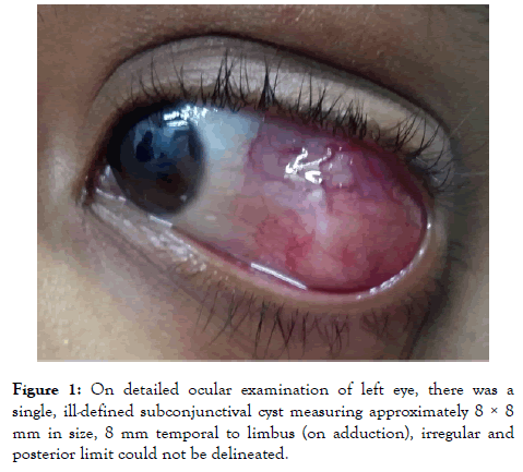
Figure 1. On detailed ocular examination of left eye, there was a single, ill-defined subconjunctival cyst measuring approximately 8 × 8 mm in size, 8 mm temporal to limbus (on adduction), irregular and posterior limit could not be delineated.
On Ultrasound Bscan orbit of left eye using a water bath, a welldefined cystic lesion measuring 7.87 mm vertically and 8.70 mm horizontally with a hyperechoic high intensity spike on upper wall of the cyst suggestive of cysticerosis cyst with scolex (Figure 2).
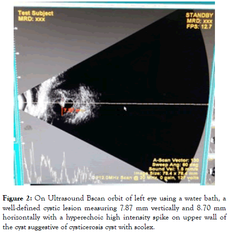
Figure 2. On Ultrasound Bscan orbit of left eye using a water bath, a well-defined cystic lesion measuring 7.87 mm vertically and 8.70 mm horizontally with a hyperechoic high intensity spike on upper wall of the cyst suggestive of cysticerosis cyst with scolex.
ELISA test of taenia solium showed positive value of 1.18. On haematological investigations, haemoglobin was 13.2 gm%, total leucocyte count was 8000 per cumm (80% polymorphs, 20% lymphocytes) and platelet count was 3.0 lac per cumm. Stool examination did not show any ova or cyst.
MRI brain with orbit confirmed diagnosis of cysticercosis with presence of eccentrix scolex associated with edema of left lateral rectus muscle with no intracranial involvement (Figure 3).
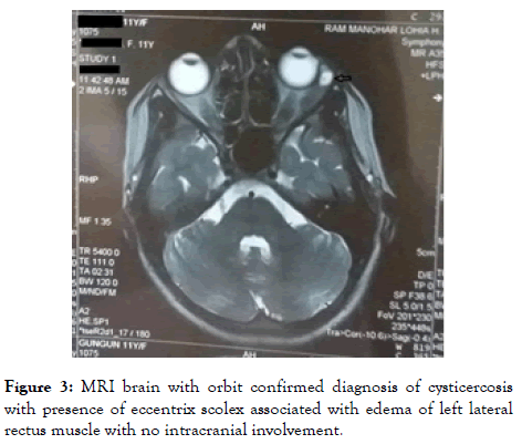
Figure 3. MRI brain with orbit confirmed diagnosis of cysticercosis with presence of eccentrix scolex associated with edema of left lateral rectus muscle with no intracranial involvement.
Patients were started on oral prednisolone–1 mg/kg per day for 7 days with gradual tapering for next 21 days. After two days of oral steroids administration, albendazole 400 mg HS was started for 21 days.
As the patient showed no improvement, cyst excision was performed under general anaesthesia. Patient was taken under general anaesthesia. Cyst was removed in toto taking care that lateral rectus muscle was not injured (Figure 4). After removal a thorough saline wash was done. The sample was send to histopathology. Post operatively topical combination of antibiotic and steroid was prescribed with gradual tapering dose over 1 month.
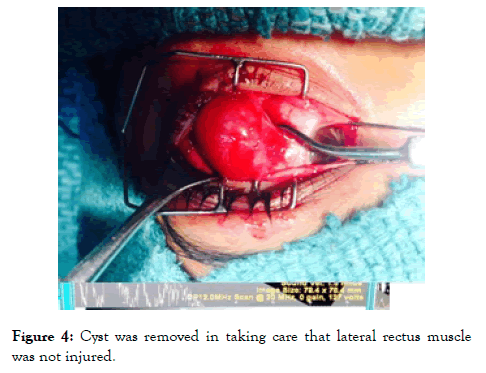
Figure 4. Cyst was removed in taking care that lateral rectus muscle was not injured.
On histopathology, section shows fibro connective along with dense mixed inflammatory infiltrate comprising of mainly lymphocytes, eosinophils, few neutrophils and plasma cells suggestive of parasitic infestation (Figures 5 and 6). No scolex or hooklets were seen probably because albendazole and steroids were given to the patient pre operatively.
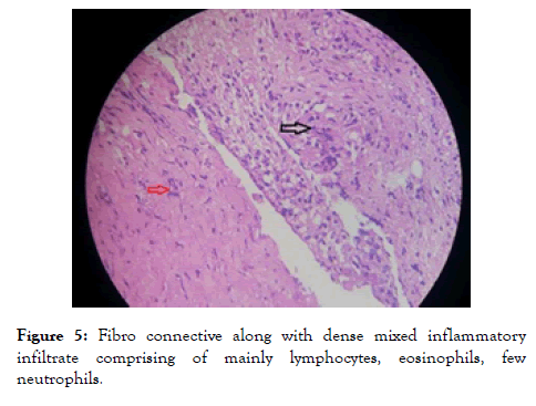
Figure 5. Fibro connective along with dense mixed inflammatory infiltrate comprising of mainly lymphocytes, eosinophils, few neutrophils.
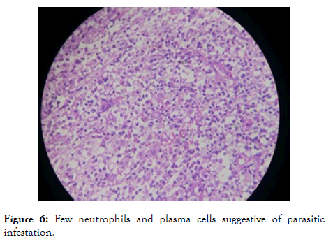
Figure 6. Few neutrophils and plasma cells suggestive of parasitic infestation.
Patient became asymptomatic with full recovery of all extraocular movements.
Discussion
Human cysticercosis occurs when Taeina solium eggs are ingested via fecal-oral transmission from a tapeworm host. Ocular cysticercosis is endemic in tropical areas including India [5]. The incidence of ocular involvement varies from 10%-30% in endemic areas. It affects individuals aged 10-30 years [6].
Subconjunctival cysticercosis usually presents as a painful, yellowish, nodular subconjunctival mass with surrounding conjunctival congestion. Acquired strabismus, diplopia, recurrent redness, and painful proptosis are some of the clinical signs in patients with orbital cysticercosis. One or more extraocular muscles may be simultaneously involved [7]. Hence detailed ocular examination followed by Serum ELISA for anticysticercal antibodies and imaging should be performed to make a final diagnosis [8].
Surgical removal remains the gold standard treatment modality for subconjunctival and eyelid cysticercosis. However cysts deep within the orbit are best treated conservatively with a 4-week regimen of oral albendazole (15 mg/kg/d) with oral steroids (1.5 mg/kg/d) in a tapering dose over a 1-month period [9]. Concomitant administration of corticosteroids is recommended to avert an inflammatory response [10].
Conclusion
Cysticercosis must be kept as a differential diagnosis of any inflammatory swelling of subconjunctival space. Meticulous systemic examination should be done as Taenia solium spreads haematogenously and spread to various organs like brain, eyes, heart and spine. A definitive diagnosis can be made by ELISA for cysticercal antibodies and imaging techniques (USG B scan and MRI). Early treatment of the subconjunctival cyst should be done as it can release toxins and involve one or more extraocular muscles leading to diplopia.
REFERENCES
- Dhiman R, Devi S, Duraipandi K, Chandra P, Vanathi M, Tandon R, et al. Cysticercosis of the eye. Int J Ophthalmol. 2017; 10(8):1319-1324.
- Kamali NI, Huda MF, Srivastava VK. Ocular cysticercosis causing isolated ptosis: A rare presentation. Ann Trop Med Public Health. 2013;6:303-305.
- Prasad KN, Prasad A, Verma A, Singh AK. Human cysticercosis and Indian scenario: A review. J Biosci. 2008; 33: 571-582
- Foyaca-Sibat H, Salazar-Campos M, Iban ez-Valde SL. Cysticercosis Of the extraocular muscles. Our experience and review Of the medical literature. The Internet J Neurol. 2012; 14(1).
- Singh RY. Subconjunctival cysticercus cellulosae. Indian J Ophthalmol. 1993; 41 : 188-189.
- Pushker N, Bajaj MS, Betharia SM. Orbital and adnexal cysticercosis. Clin Exp Ophthalmol. 2002 ; 30(5):322-333.
- Das S. Anterior orbital cysticercosis: A case presentation. Kerala J Ophthalmol. 2017; 29:230-233.
- Magu S, Nada M, Khurana AK, Chugh JP. Spontaneous extrusion of subconjuctival cysticercus cellulosae. Br J Ophthalmol. 2001; 85(1):116-117.
- Rai PJ, Nair AG, Trivedi MG, Potdar NA, Gopinathan I, Shinde CA. An unusual eyelid mass of cysticercosis: A twist in the tale. J Cutan Aesthet Surg. 2016; 9(2):126-128.
- Kaliaperumal S, Rao VA, Parija SC. Cysticercosis of the eye in south India: A case series. Indian J Med Microbio. 2005; 23:227-230.
Citation: Ish S, Pathak A, Sharma R, Hasan S (2019) An Interesting Case of Subconjunctival Cysticercus Cyst: A Case Report. J Eye Dis Disord. 4:129. DOI: 10.35248/ 2684-1622.19.4.129
Copyright: © 2019 Ish S, et al. This is an open-access article distributed under the terms of the Creative Commons Attribution License, which permits unrestricted use, distribution, and reproduction in any medium, provided the original author and source are credited.
Sources of funding : No Funding
