Indexed In
- RefSeek
- Hamdard University
- EBSCO A-Z
- Euro Pub
- Google Scholar
Useful Links
Share This Page
Journal Flyer
Open Access Journals
- Agri and Aquaculture
- Biochemistry
- Bioinformatics & Systems Biology
- Business & Management
- Chemistry
- Clinical Sciences
- Engineering
- Food & Nutrition
- General Science
- Genetics & Molecular Biology
- Immunology & Microbiology
- Medical Sciences
- Neuroscience & Psychology
- Nursing & Health Care
- Pharmaceutical Sciences
Case Report - (2019) Volume 4, Issue 1
Acquired Tufted Angioma: A Clinicopathological Entity
Manisha Nijhawan1, Shivi Nijhawan1, Kingshuk Chatterjee2, Govind srivastava3 and Virendra N Sehgal4*2Department of Dermatology, Calcutta School of Tropical Medicine, West Bengal, India
3Skin Institute and School of Dermatology, Greater Kailash, New Delhi, India
4Department of Dermato Venereology (Skin/VD) Center, Sehgal Nursing Home, Panchwati-Delhi, India
Received: 04-May-2018 Published: 09-Jan-2019
Abstract
Acquired angioma, an asymptomatic clinicopathologic entity derived from cells of the vascular or lymphatic vessel walls and the lymphatic wall tissues surrounding these vessels, a significant benign skin marker, tufted angioma being one of them wherein its histopathology is characterized by multiple circumscribed round or ovoid vascular tufts and lobules of densely packed capillaries, randomly scattered throughout the mid, lower dermis and subcutaneous fat in a typical “Cannon Ball Pattern”, the relevant literature has been reviewed in brief.
Keywords
Vascular tumors; Acquired; Atypical
Introduction
Acquired angiomas, vascular tumors perse has thus far been sporadically reported [1,2] because of its asymptomatic nature despite explicit clinicopathological features. The current report is an attempt to focus attention on these aspects, enriching through clinical as well as histological illustrations. A 30-year-old woman presented with reddish, nodular lesions scattered around the left eye, (Figure 1) for the past 5 years. The initial lesion appeared over the medial canthus, which was excised 3 years ago but there was a recurrence at the same site, and also at upper lateral border of left eyebrow, accompanied by mild tenderness. There was neither a history of trauma, bleeding nor any systemic complains. Hematoxylin and Eoisin (H&E) stained skin section(s) depicted moderate hyperkeratosis, acanthosis and elongation of rete ridges (Figure 2) in addition to numerous dilated, simulating cannon ball (Figure 3) thin and thick walled capillaries within the dermis.
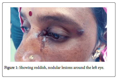
Figure 1. Showing reddish, nodular lesions around the left eye.
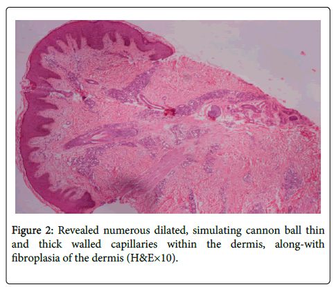
Figure 2. Revealed numerous dilated, simulating cannon ball thin and thick walled capillaries within the dermis, along-with fibroplasia of the dermis (H&E×10).
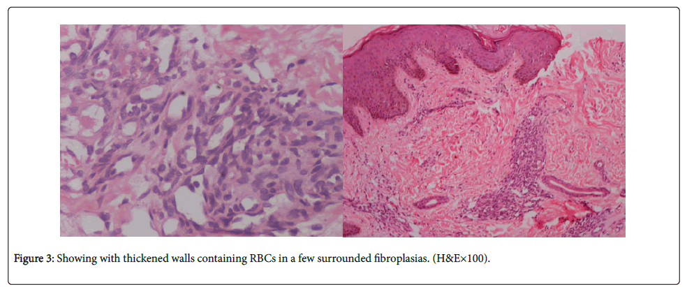
Figure 3. Showing with thickened walls containing RBCs in a few surrounded fibroplasias. (H&E×100).
The lining of the capillaries was of plump or elongated endothelial cells. Some of the capillaries had thickened walls and were engorged with red blood cell (RBCs). The dermis surrounding these clusters showed fibroplasia (Figure 3). Mild to moderate hyperplasia of the epidermis was a concomitant feature. This particular patient prompted to look for such cases in the course the following 5 years, accordingly, we succeeding in identifying 4 other clinical variants arising from cells of the vascular or lymphatic vessel walls and the lymphatic wall tissues surrounding these vessels (Table 1). The salient clinical and histological finding of which are succinctly portrayed in the adjoining table (Table 1).
| Serial No. | Age in years(y) and Gender(M/F) | Clinical presentation | Histopathology feature | Diagnosis |
|---|---|---|---|---|
| Case 1 | 28/F | Index case | - | Acquired tufted angioma |
| Case 2 | 35/F | Multiple, hyper keratotic nodular lesions of U/L configuration, distributed on the right upper extremity of 2 years’ duration. | H & E stained section(s) of the skin depicted diffuse, dense vascular proliferation of thick walled capillaries, and solid endothelial cords with abundant extravasation of RBCs. The surface was covered by fibrin and necrotic cells. The stroma showed numerous neutrophils and edema. A few thickened bundles of collagen were found to traverse the lesion, suggestive of septa formation. The Grams’ stain was negative for microorganisms. | Multiple pyogenic granuloma |
| Case 3 | 30/M | Asymptomatic, reddish nodular lesions affecting the right ear of 7 month’s duration. | H & E stained section(s) of the skin showed an increased number of thick walled and dilated capillaries and venules, involving the whole of reticular dermis. An infiltrate of lymphocytes and a few eosinophils were conspicuous around these vessels and occasionally seen infiltrating their walls. | Angiolymphoid hyperplasia |
| Case 4 | 35/M | Pin-head to pea size reddish-purple, raised and grouped lesions distributed over the left upper arm of 8 months duration. | H & E stained section(s) of the skin depicted a well-circumscribed proliferation of numerous thin walled dilated capillaries, displaying a nodular pattern along the superficial-and deep plexuses. Most of them showed a complete lining of endothelial cells, along with abundant hemosiderin deposits around them. A few thick walled capillaries with projecting endothelial cells were also seen. Hemosiderin deposits were confirmed by using prusian blue stain. | Hemosiderotic hemangioma |
Table 1: Portraying clinical and histological findings of other than index case.
The study of angioma is a fascinating overture, ever since its recognition as a clinical entity in the year 1976 by E. Wilson-Jones [1,2] it is therefore, incumbent to define angioma [3] in order to comprehend its evolution in perspective. Angiomas are benign tumors, derived from cells of the vascular or lymphatic vessel walls and the lymphatic wall tissues surrounding these vessels. Angiomas are a significant marker of age but may also be an expression of systemic disorder, the portal cirrhosis manifesting as caput medusae, the palm tree sign, an appearance of distended and engorged superficial epigastric veins, radiating from the umbilicus across the abdomen. Usually they are benign and are not associated with malignancy [4,5]. Most often access to specialist is because of cosmetic abrasion. Its histopathology is pathognomonic [1,6] characterized by Multiple circumscribed round or ovoid vascular tufts and lobules of densely packed capillaries randomly scattered throughout the mid and lower dermis and subcutaneous fat in a typical “Cannon Ball Pattern”.
The aggregates of endothelial cells form a concentric whorl along a pre-existing vascular plexus.
Some lobules bulge the walls of dilated thin walled vascular structures, giving a semi-lunar appearance to the vessels.
Immunohistochemically, the cells in the capillary tufts are positive for CD31, CD34 and smooth muscle actin.
Histopathology, therefore, is warranted to be carried in all cases of angioma. However, it is required to be differentiated form capillary hemangiomas, a congenital abnormality [7], by and large are benign tumor emanating from the blood vessel(s), primarily made up of blood vessels, may either be capillary, Lobular capillary (lobuler capillary of pregnancy), cavernous or compound. Capillary hemangiomas have a distinct clinical presentation looking like red-wine and strawberrycolour may protrude from the skin.
Diascopy, (blanchability) test is performed by applying pressure with a finger. It is a useful office procedure. They appear most frequently on the face, neck, and behind the ears and are largely asymptomatic.
Conclusion
Currently, angioma an up front, identified as an entity in the year 1976 it reporting since then has been sporadic.
Angiomas are benign tumors, derived from cells of the vascular and/or lymphatic vessel walls.
Angiomas by and large are asymptomatic, Predominantly affects the upper part of the body.
Multiple lobules of poorly canalized capillary channels as “Cannon balls” in dermis and subcutis are histopathological hallmarks of the disease.
Cosmetic reason may induce for concentration, Although spontaneous regression has been seen to occur within 6 months to 2 years, local recurrences have warranted surgical interventions.
Nonetheless, morphology of the lesions complemented by pathogonomic histopathology is diagnostic.
Clinical as well as histopathological features therefore formed a subject of focus in order to create awareness about the prevalence entity. Anticipating similar reports in the near future.
REFERENCES
- Descours H, Grézard P, Chouvet B, Labeille B (1998) Acquired tufted angioma in an adult. Ann Dermatol Venereol 125: 44-46.
- Hebeda CL, Scheffer E, Starink TM (1993) Tufted angioma of late onset. Histopathology 23: 191-193.
- https://medical-dictionary.thefreedictionary.com/angioma.
- James WD, Berger TG, Elston DM, Odom RB (2006) Andrews' Diseases of the Skin: Clinical Dermatology, Philadelphia, Saunders Elsevier, United States.
- Rapini RP, Bolognia JL, Jorizzo JL (2007) Dermatology: 2-Volume Set. St. Louis: Mosby. pp: 1779
- Requena L, Sangueza OP (1997) Cutaneous vascular proliferation. Part II. Hyperplasias and benign neoplasms. J Am Acad Dermatol 37: 887-919.
- Macmillan A, Champion RH (1971) Progressive capillary haemangioma. Br J Dermatol 85: 492-493.
Citation: Nijhawan M et al. (2019) Acquired Tufted Angioma: A Clinicopathological Entity. J Dermatitis 4:115.
Copyright: © 2019 Nijhawan M, et al. This is an open-access article distributed under the terms of the Creative Commons Attribution License, which permits unrestricted use, distribution, and reproduction in any medium, provided the original author and source are credited.
