Indexed In
- The Global Impact Factor (GIF)
- CiteFactor
- Electronic Journals Library
- RefSeek
- Hamdard University
- EBSCO A-Z
- Virtual Library of Biology (vifabio)
- International committee of medical journals editors (ICMJE)
- Google Scholar
Useful Links
Share This Page
Journal Flyer

Open Access Journals
- Agri and Aquaculture
- Biochemistry
- Bioinformatics & Systems Biology
- Business & Management
- Chemistry
- Clinical Sciences
- Engineering
- Food & Nutrition
- General Science
- Genetics & Molecular Biology
- Immunology & Microbiology
- Medical Sciences
- Neuroscience & Psychology
- Nursing & Health Care
- Pharmaceutical Sciences
Case Report - (2024) Volume 23, Issue 3
A singular approach of ridge preservation: Socket shield technique.
Abhishek Singh*Received: 22-Sep-2020, Manuscript No. OHDM-24-6560; Editor assigned: 25-Sep-2020, Pre QC No. OHDM-24-6560 (PQ); Reviewed: 09-Oct-2020, QC No. OHDM-24-6560; Revised: 02-Sep-2024, Manuscript No. OHDM-24-6560 (R); Published: 30-Sep-2024, DOI: 10.35248/2247-2452. 24.23.1116
Abstract
With the aim of obtaining an excellent aesthetic result, implant dentistry has turn into a prosthetically driven practice. The socket shield technique has exhibited the possibility in avoiding buccal tissue from resorption in animal and clinical studies. In order to reduce the resorption of hard and soft tissue, which prevails around the newly extracted tooth to form a natural emergence profile, socket preservation technique was brought in. It is pretended that retaining a root remnant adhered to the buccal bone plate can evade tissue change after tooth extraction. This case report depicts of a young patient with fractured tooth in left central incisor, in which immediate implant placement with socket shield technique was initiated where root was bisected and buccal two-third of the root was retained in the socket so that the periodontium along with the bundle bone and buccal bone remains intact.
Keywords
Extraction socket, Buccal tooth fragment, Tooth retention, Ridge preservation, Socket shield, Immediate implant placement, Synthetic bone graft, Osseointegration.
Introduction
One of the primary objective of prosthetic rehabilitation is to attain harmony between the pink and white zones especially in the esthetic areas. Various researches confirm that buccal part of the ridge is more compromised as it is chiefly supplied by periodontal membrane of the tooth. After tooth extraction, buccal cortical plate starts faster resorption than palatal/lingual plate which may lead to partial or total resorption [1].
To overcome these adverse effects of tooth extraction, various methods have been reported in the literature such as hard and soft tissue augmentation following extraction with or without immediate implant placement. Socket shield technique was first described in 2010 aimed at leaving the buccal fragment of root intact and placing implant on the lingual aspect of that fragment so that the tissues which remains in contact with the buccal fragment hold its vitality and avoid ridge collapse.
This case report describes a patient whose alveolar ridge of a fractured tooth is preserved by "socket shield technique" which can prove to be helpful for implantologists when planning implants in aesthetic zone [2].
Procedure
The steps of socket shield technique used for immediate implant placement are outlined as:
Step 1: Cut the crown horizontally at the gingival level (Figure 1).
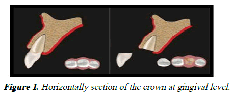
Figure 1: Horizontally section of the crown at gingival level.
Step 2: Bisect the root vertically in such a way that palatal half is detached along with the apex. The length of the shield should be kept at two‑third of the root length and buccal part is then reshaped such that the shield width is about 1.5 mm-2.0 mm. The shield should be trimmed to the bone level. A bevel or Sshaped profile on the inner side of the shield is given to hold the restorative segment (Figure 2) [3].
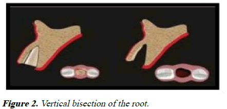
Figure 2: Vertical bisection of the root.
Step 3: Placement of implant in correct three‑dimensional area. The optimal space between shield and implant is 1.5 mm or more. A bone graft is implied if the gap is more than 3.0 mm. An interim crown or a customized healing abutment given instantly after the implant placement that will help in preserving the soft tissue contours. The choice of prosthesis for the final restoration is a screw-retained crown or a cement-retained crown with restorative margin that can be well accessed for cement clean up (Figure 3).
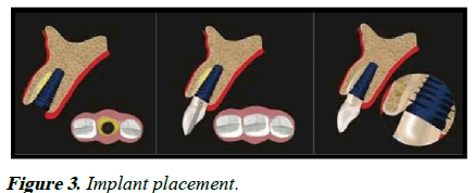
Figure 3: Implant placement.
The principle of socket shield technique is as follows:
• Preparation of the root of a tooth indicated for extraction in
such way that the buccal/facial root part remains situated to
the buccal plate intact.
• The tooth root section’s periodontal attachment stands vital
and intact to restrict post-extraction socket remodeling and
to hold the buccal/facial tissues.
• The prepared tooth root part acts as a socket shield and
inhibits the inflation of tissues buccofacial to an
immediately placed implant [4].
Socket shield technique was first defined by Hurzeler et al in 2010. The procedure consists of leaving a root fragment when extracting the tooth, specially the vestibular section of the most coronal third of the root. The socket shield technique is aimed at making up for loss of the vestibular volume, the bundle bone because the periodontal ligament remains adhered to the dentin and cementum of the root fragment. Various animal studies exhibited that the loss of volume after extraction could vastly decrease when leaving a tooth portion adhered to the cortical bone in the vestibular part of the alveolus. The socket shield technique is still missing clinical long-term statistics to be favored as a usual therapy.
Case Presentation
A young male patient came for the treatment of a fractured left central incisor (Figures 4).
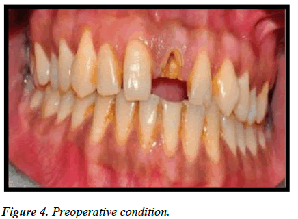
Figure 4: Preoperative condition.
The medical history was not revealed. Implant placement with socket shield technique was preferred as treatment of choice. After subsequent application of local analgesia, the tooth was sectioned at gingival level and then divided into buccal and palatal parts using a long root resection bur (Figures 5 and 6) [5].
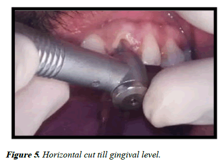
Figure 5: Horizontal cut till gingival level.
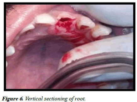
Figure 6: Vertical sectioning of root.
It was designed to retain the buccofacial half of the root intact. Periotomes were used to split the periodontal ligament and palatal section of root was precisely detached without harming the buccal root section (Figure 7).
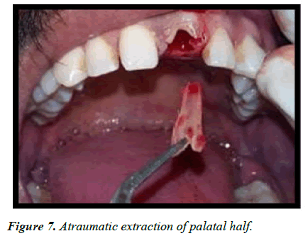
Figure 7: Atraumatic extraction of palatal half.
The remaining root segment was then carved properly and shortened coronally to the level of alveolar crest. The frame of the section was done by trimming it both in apicocoronal and mesiodistal direction (using a long-shanked, large and round diamond bur). The extraction socket was then curetted to eliminate all granulation tissues and buccal root shield was tested for immobility by applying a sharp probe to its surface. Once perfectly adapted, this root section is noted as socketshield or root membrane. The implant placement step was done as per the drilling order implied by the implant manufacturer. The drilling was proposed using a lance drill to employ the palatal aspect of the root so that the buccal aspect would stay intact. Ensuing implant bed preparation, a tapered internal hex implant, 3.8 mm × 15 mm was arranged in correct 3D position (Figures 8 and 9) [6].
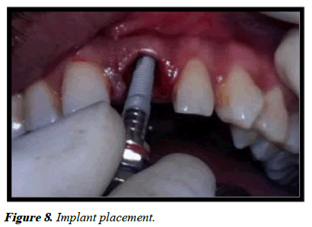
Figure 8: Implant placement.
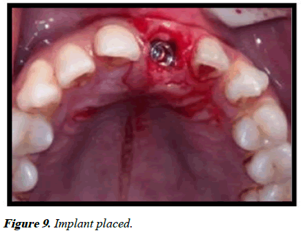
Figure 9: Implant placed.
The implant so placed had mesial, distal and palatal bony walls. On the buccal side, it had the remaining buccal section of the root that had thin layer of dentin, followed by cementum, periodontal ligament and bundle bone in socket-facial direction. After implant placement, a screw-retained temporary crown was made for immediate implant placement in the esthetic region (Figures 10) [7].
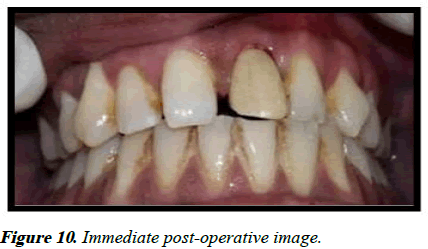
Figure 10: Immediate post-operative image.
Following fabrication of the interim restoration, a thorough occlusal review was achieved to establish nonfunctional loading. Postsurgical instruction includes antibiotics and analgesic aid and chlorhexidine 0.12% oral rinse. The patient was further advised to hold up from tooth brushing or any mechanical injury in the area for 2 weeks. After 2 weeks, the patient is asked to get for a checkup. Clinical and radiographic interpretation of the site was done after 3 months postoperatively. Complete preservation of hard and soft tissue was recorded at the surgical site. The usual clinical protocol was used for the fabrication of the definite restoration. Zirconia abutments were used to contrive the esthetically favorable lithium disilicate crowns (Figures 11).
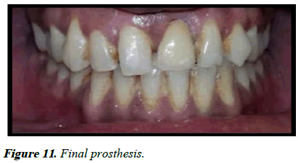
Figure 11: Final prosthesis.
The patient was kept on follow‑up 3 months, 6 months and yearly thence-forth [8].
Discussion
Apart from aesthetics, alveolar ridge atrophy after tooth extraction has a negative impact on implant supported prosthetic restoration. Various methods have been reported in the literature such as immediate implant, bone grafts and membranes have been used in this view. However, immediate implant placement still does not inhibits buccal bone resorption as it is a biological aspect. Some studies show that de-crowned root fragments not only adequately retains bone volume but also creates vertical growth in coronal direction. Ridge preservation is of absolute concern if good diagnosis is needed. This case report offers one such method i.e., the socket shield technique which defines that by preserving the facial fragment of the root, thin labial cortical bone and contour of the ridge is retained. Histological studies have shown new bone formation in the small gap between implant in contact with the tooth fragment. However, a layer of new cementum was found between implant and tooth portion when enamel matrix derivative (emdogain) was enforced to the inner surface of the tooth fragment before implant placement. So, in this case report it was shown that socket shield technique with immediate implant placement preserves the buccal cortical plate and normal peri-implant tissue. Still this technique with its specific upcoming outcome, will shortly be included as a routine practice in ridge preservation [9].
Advantages of socket shield technique
It is a minimally invasive surgical procedure, which not only preserves a part of the tooth but also helps to maintain hard and soft tissue contours, as long as the shield is intact. It shortens the need of soft and hard tissue grafting measures, interdental papilla can be preserved by preparing interdental socket shield and minimizes the overall treatment period. This is a well promising technique in terms of managing pink and white esthetics and brings a solution for esthetically demanding cases such as maxillary anteriors and high lip line.
Limitations of socket shield technique
The clinician needs to be specially trained and to have high level of clinical skills. If the shield becomes mobile during surgery, it is removed and conventional immediate implant placement or grafting procedure is to be done. Case selection is very necessary for the success of the procedure. This approach is not preferred in mobile teeth, teeth which are out of the arch and teeth with large periapical lesions. The intactness of the shield plays a basic role in the success of the treatment [10].
Conclusion
The socket shield technique is getting popularity among the clinicians across the world. The technique is very favorable for the preservation of hard and soft tissues in cases of post extraction immediate implant placement. In selected cases, immediate placement of implants with socket shield technique looks a suitable means for the replacement of lost teeth, chiefly in the aesthetic area. Yet histological data and long term pursue has to be directed to confirm socket shield technique on routine base. It can be concluded that retaining buccal aspect of the root in combination with immediate implant placement is a feasible technique to obtain osseointegration without any inflammatory or resorptive reply.
References
- Hurzeler MB, Zuhr O, Schupbach P, Rebele SF, Emmanouilidis N, Fickl S. The socket‐shield technique: A proof‐of‐principle report. J Clin Periodontol. 2010;37(9):855-862.
[Crossref] [Google Scholar] [PubMed]
- Baumer D, Zuhr O, Rebele S, Schneider D, Schupbach P, Hurzeler M. The socket‐shield technique: First histological, clinical and volumetrical observations after separation of the buccal tooth segment-a pilot study. Clin Implant Dent Relat Res. 2015;17(1):71-82.
[Crossref] [Google Scholar] [PubMed]
- Mitsias ME, Siormpas KD, Kotsakis GA, Ganz SD, Mangano C, Iezzi G. The root membrane technique: Human histologic evidence after five years of function. Biomed Res Int. 2017;2017(1):7269467.
[Crossref] [Google Scholar] [PubMed]
- Kan JY, Rungcharassaeng K. Proximal socket shield for interimplant papilla preservation in the esthetic zone. Int J Periodontics Restorative Dent. 2013;33(1):e24-31.
[Crossref] [Google Scholar] [PubMed]
- Lee CT, Chiu TS, Chuang SK, Tarnow D, Stoupel J. Alterations of the bone dimension following immediate implant placement into extraction socket: Systematic review and meta‐analysis. J Clin Periodontol. 2014;41(9):914-926.
[Crossref] [Google Scholar] [PubMed]
- Glocker M, Attin T, Schmidlin PR. Ridge preservation with modified “socket-shield” technique: A methodological case series. Dent J. 2014;2(1):11-21.
- Filippi A, Pohl Y, Von Arx T. Decoronation of an ankylosed tooth for preservation of alveolar bone prior to implant placement. Dent Traumatol. 2001;17(2):93-95.
[Crossref] [Google Scholar] [PubMed]
- Elad A, Rider P, Rogge S, Witte F, Tadic D, Kacarevic ZP, et al. Application of biodegradable magnesium membrane shield technique for immediate dentoalveolar bone regeneration. Biomedicines. 2023;11(3):744.
[Crossref] [Google Scholar] [PubMed]
- Chu SJ, Tan-Chu JH, Levin BP, Sarnachiaro GO, Lyssova V, Tarnow DP. A paradigm change in macro implant concept: Inverted body-shift design for extraction sockets in the esthetic zone. Compendium. 2019;40(7):444-452.
[Google Scholar] [PubMed]
- Kunrath MF, Gerhardt MD. Trans-mucosal platforms for dental implants: Strategies to induce muco-integration and shield peri-implant diseases. Dent Mater. 2023;39(9):846-859.
[Crossref] [Google Scholar] [PubMed]
Citation: Singh A. A singular approach of ridge preservation: Socket shield technique. Oral Health Dent Manage. 2024;23(3):1116.
