Indexed In
- Open J Gate
- Genamics JournalSeek
- Academic Keys
- JournalTOCs
- ResearchBible
- China National Knowledge Infrastructure (CNKI)
- Scimago
- Ulrich's Periodicals Directory
- Electronic Journals Library
- RefSeek
- Hamdard University
- EBSCO A-Z
- OCLC- WorldCat
- SWB online catalog
- Virtual Library of Biology (vifabio)
- Publons
- MIAR
- Scientific Indexing Services (SIS)
- Euro Pub
- Google Scholar
Useful Links
Share This Page
Journal Flyer

Open Access Journals
- Agri and Aquaculture
- Biochemistry
- Bioinformatics & Systems Biology
- Business & Management
- Chemistry
- Clinical Sciences
- Engineering
- Food & Nutrition
- General Science
- Genetics & Molecular Biology
- Immunology & Microbiology
- Medical Sciences
- Neuroscience & Psychology
- Nursing & Health Care
- Pharmaceutical Sciences
Research Article - (2023) Volume 14, Issue 2
: Ascaridia galli (chicken nematode) as a Source for Nanoparticle and Nanofiber
Sunila Kumari*Received: 03-Feb-2023, Manuscript No. JNMNT-23-19633; Editor assigned: 06-Feb-2023, Pre QC No. JNMNT-23-19633; Reviewed: 20-Feb-2023, QC No. JNMNT-23-19633; Revised: 24-Feb-2023, Manuscript No. JNMNT-23-19633; Published: 28-Feb-2023, DOI: 10.35248/2157-7439.23.14.663.
Abstract
Green synthesis of nano particles and nanofiber gained relevance in recent times. Ideally nanoparticles and nanofiber are made from metallic sources. However, in recent times, there are reports of drastic side effects about the use of metallic nanoparticles in biology and medicine. In this context, it has become more relevant to explore and identify, more natural and biofriendly and effective alternative to metallic ones. There are instances, where, nanoparticles and nanofibers, and many other nano components, have been synthesized from more natural sources, such as bacteria, different species of algae and also from fungi. However, not many have attempted to synthesize nanoparticles, from unusual sources such as nematodes, the intestinal parasites of animals and humans. For the first time, in the present study it was attempted to synthesize nanoparticles and nanofiber from the intestinal nematode of chicken, Ascaridia galli. The parasites, were extracted and isolated from the chicken intestines, they were later processed for the synthesis of nanoparticle and nanofiber. It is quite evident that nanoparticles, synthesized from a natural source are more appealing as they are both ecofriendly and inexpensive. Parasite, measuring approximately 4 to 5cms in length were carefully removed from chicken intestines and later processed for the synthesis of nanoparticle and nanofiber. Nanoparticles were synthesized using the method of acid hydrolysis and nanofiber was prepared using electro spinning technique. The nanofibre which was synthesized was hydrophilic in nature. Scanning electron microscope (SEM) images of nanoparticle revealed a diameter of 48.46nm- 20μm and the nanofiber diameter was found to be between 88nm-2 μm, thus matching to the average standard size of nano particle and nanofiber. As the intestinal parasites, are capable of evading host immunity and can easily amalgamate with the host tissues, without triggering any antagonism, therefore can be used frequently in biology and medicine. As these nanoparticles and nanofiber, are synthesized using a parasite,(a living source) it can easily be degraded after the use, and may be considered as a suitable alternative to metallic nanomaterials.
Keywords
Nanoparticle; Nanofiber; Ascaridia galli; Acid hydrolysis, Electro spinning; SEM; Chicken nematode
Introduction
In recent times, nano technology has its impacted almost every walks of life. Its importance is ever growing in the field of medicine and regenerative biology. Nanoparticle, nanomembranes, nanofibers, nanorods, find applications in almost every walk of life, ranging from, industrial, biological, pharmacological and biomedical fields [1]. The unique versatility, of nanoparticles, is attributed to their, size and dimensions (100 nm) and it is interesting to note that materials, exhibit certain unique physical properties, when they are downsized to nano proportions, in contrast to heavier materials, which show constant physical properties [2]. The primary cause for the development of nanotechnology, stems, from the understanding of biological processes as they are often happening at a nanoscale [3] and living cells are typically 10μm in size and their components are much smaller in size. Metals and heavy metals are routinely used for the synthesis of nanoparticles, which are usually employed in many biological and medical applications. In spite of being used in biological and medical applications, they reportedly have deleterious effects upon human body [4]. In order to minimize or to totally eliminate the risk of side effects, there are attempts to constantly explore and identify different materials, which can be both biologically friendly and also cost effective. In this regard, a number of plant based nanoparticles, have been synthesized and this method of developing plant based nanoparticles, is considered to be green chemistry approach [5]. Apart from plant materials, being used as nanoparticles, there are evidences, where in microorganism, such as bacteria, fungi yeast are also used to synthesize nano products [6]. Cellulose and bacterial cellulose (BC) seem to be used for synthesis of ecofriendly, nano based products, which seem to gain more popularity in recent times [7,8]. Therefore from these studies it is very much obvious that nanoparticles, synthesized from natural sources are more appealing as they are biocompatible and also least expensive. Therefore present study is an attempt to develop nanoparticles and nanofiber from much cheaper source or to be precise, from waste material i.e. from an intestinal parasite, chicken nematode, and Ascaridia galli.
RESULTS
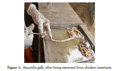
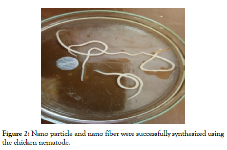
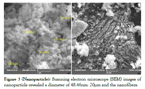
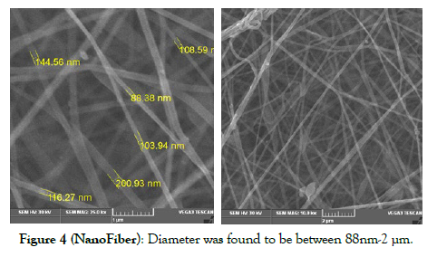
Nano particle and nano fiber were successfully synthesized using the chicken nematode, (Figure 2) Ascaridia galli, after being extracted from chicken intestines (Figure 1). Scanning electron microscope (SEM) images of nanoparticle (Figure 3) revealed a diameter of 48.46nm- 20μm and the nanofiber (Figure 4) diameter was found to be between 88nm-2 μm. These measurements are in accordance with the average standard size of nanoparticle and nanofiber, which are routinely used in scientific and other biological applications.

Figure 1: Ascaridia galli, after being extracted from chicken intestines.

Figure 2: Nano particle and nano fiber were successfully synthesized using the chicken nematode.

Figure 3: Scanning electron microscope (SEM) images of nanoparticle revealed a diameter of 48.46nm- 20μm and the nanofibers.

Figure 4: Diameter was found to be between 88nm-2 μm.
MATERIALS AND METHOD
For the purpose the experimentation, chicken intestines, were collected from the source. The intestines, infested with parasitic infection, were thoroughly washed, and then dissected for parasitic infection, chicken nematode, identified as Ascaridia galli, was identified, which was white in color and was carefully collected in to the petridish. The worms, measured approximately 4-5 cms in length. They were collected and dried overnight in an incubator at a temperature of 37 degree Celsius. The worms after thoroughly being dried were pounded into a fine powder using a motor and pestle, and were further processed for the synthesis of nanoparticles and nanofiber. Scanning electron Microscope (SEM) images were taken to ascertain the size of nanofiber and nanoparticle.
Preparation of Nanoparticles
Nematode nanoparticles were produced from the purified nematode powder by repeated acid hydrolysis. Nematode powder was soaked in 3M HCl for acid hydrolysis and incubated for 90 min at 90°C in a thermostat water bath. The sample was centrifuged at 6000 rpm for 10 min and the pellets were collected. The acid hydrolysis step was repeated thrice and the final pellet was suspended in distilled water to dilute the acid concentration. The suspension was dialyzed against distilled water until it reaches pH 6.
Preparation of Nanofibre
Nematode powder of 2% was dissolved in 80 %( w/v) of acetic acid. To the mixture 1 ml of 10% PVA (Poly vinyl alcohol) polymer was added as additive to obtain thick fibre consistency. The mixture was electro spinned in 18kv potential using electro spinning apparatus, and tip to plate distance of 15 cm at a flow rate of 0.5 ml/hour
DISCUSSION
In the present study, I have successfully synthesized nanoparticles and a hydrophilic nanofiber, using a rather unusual source, chicken intestinal nematode, Ascaridia galli. As mentioned earlier, metals and other non-metal ions, though can be effectively molded into various nanostructures, but may not be useful to be used in medical and biological applications, because of their side effects. Nano particles are frequently used in cancer treatment because of their ability to directly deliver to the desired cells [9, 10] and also to target precisely the tumor cells amongst the healthy ones. Apart from nano particles, nanofibers are also used in tissue engineering, bone remodeling and skin regeneration. In addition to these, applications, nanofibers are also used is in wound healing and wound treatment, because of their low weight, nano scale porosity and nano diameter [11]. In a recent study by authors such as Lihua [12] suggest that nano fiber applications, seem to show, promising results, in the field of energy storage protective clothing, and air/ gas filtration. In conclusion it may be said, that, nano structures, derived from natural components, may be frequently used in biological and medical applications, as they are more ecofriendly, cost effective and more importantly biocompatible. Therefore in the present study I have successfully derived nanoparticles and nanofiber from a rather natural or unusual source, an intestinal nematode parasite.
STATEMENTS AND DECLARATIONS
Funding
No funding was provided for the project from any government or private agencies
Competing interests
Since this is a single author paper, no competing interests exist.
Ethics approval
Author, declares that no animal was harmed or injured in the process of experimentation. As the parasites were collected from the intestines, of sheep, which were collected from the source (slaughter houses) as discarded material, hence no harm was caused to any animal.
REFERENCES
- Wang EC, Wang AZ. Nanoparticles and their applications in cell and molecular biology. Integr Biol. 2014; 6(1): 9-26.
- Abdel Goad M, Mahmoud N, Saad B. The Analysis of Adsorption Phenomenon for Nano silica with Some Radionuclides Released in The Primary Coolant of PWR. Egypt J Chem. 2020; 63(5): 13-14.
- Kipp J. The role of solid nanoparticle technology in the parenteral delivery of poorly water-soluble drugs. Int J Pharm. 2004; 284(1-2): 109-122.
- Agarwal M, Murugan M, Sharma A, Rai R, Kamboj A, Sharma H, et al. Nanoparticles and its toxic effects: a review. Int J Curr Microbiol Appl Sci. 2013; 2(10): 76-82.
- Parveen K, Banse V, Ledwani L. Green synthesis of nanoparticles: their advantages and disadvantages. AIP Conf Proc. 2016; 1724: 020048
- Li X, Xu H, Chen ZS, Chen G. Biosynthesis of nanoparticles by microorganisms and their applications. J Nanomater. 2011; 1-16.
- Jose J, Thomas V, Raj A, John J, Mathew RM, Vinod V, et al. Eco‐friendly thermal insulation material from cellulose nanofibre. J Appl Polym Sci. 2020; 137(2): 48272.
- Ma L, Bi Z, Xue Y, Zhang W, Huang Q, Zhang L, et al. Bacterial cellulose: an encouraging eco-friendly nano-candidate for energy storage and energy conversion. J Mater Chem. 2020; 8(12): 5812-5842.
- Hammel E, Tang X, Trampert M, Schmitt T, Mauthner K, Eder A, et al. Carbon nanofibers for composite applications. Carbon. 2004; 42(5-6): 1153-1158.
- Verreck G, Chun I, Rosenblatt J, Peeters J, Van Dijck A, Mensch J, et al. Incorporation of drugs in an amorphous state into electrospun nanofibers composed of a water-insoluble, nonbiodegradable polymer. J Control Release. 2003; 92(3): 349-360.
- Wróblewska-Krepsztul, Jolanta, Tomasz Rydzkowski, Iwona Michalska-Pożoga, Vijay K Thakur. Biopolymers for Biomedical and Pharmaceutical Applications: Recent Advances and Overview of Alginate Electrospinning. Nanomaterials. 2019; 9(3): 404.
- Lihua Lou, Odia Osemwegie, Seshadri Ramkumar S Functional Nanofibers and Their Applications. Ind Eng Chem Res. 2020; 59 (13): 5439-5455.
Indexed at, Google Scholar, Crossref
Indexed at, Google Scholar, Crossref
Indexed at, Google Scholar, Crossref
Indexed at, Google Scholar, Crossref
Indexed at, Google Scholar, Crossref
Indexed at, Google Scholar, Crossref
Indexed at, Google Scholar, Crossref
Indexed at, Google Scholar, Crossref
Indexed at, Google Scholar, Crossref
Indexed at, Google Scholar, Crossref
Citation: Kumari S (2023) Ascaridia Galli (Chicken Nematode) As a Source for Nanoparticle and Nanofiber. J Nanomed Nanotech. 14: 663.
Copyright: ©2023 Kumari S. This is an open-access article distributed under the terms of the Creative Commons Attribution License, which permits unrestricted use, distribution, and reproduction in any medium, provided the original author and source are credited.


